Skip to main content
Fish Pathogens
Menu
Search
Search
Main navigation
Image Galleries
Pathogens
Pathogens sub-navigation
10 Common Pathogens
What's That Spot?
Ask an Expert
FAQ
Search
Search
Search
User account menu
Log in
Primary tabs
Common Pathogens
All Pathogens
Hosts
Field Sites
Videos
Contributed
All Pathogens
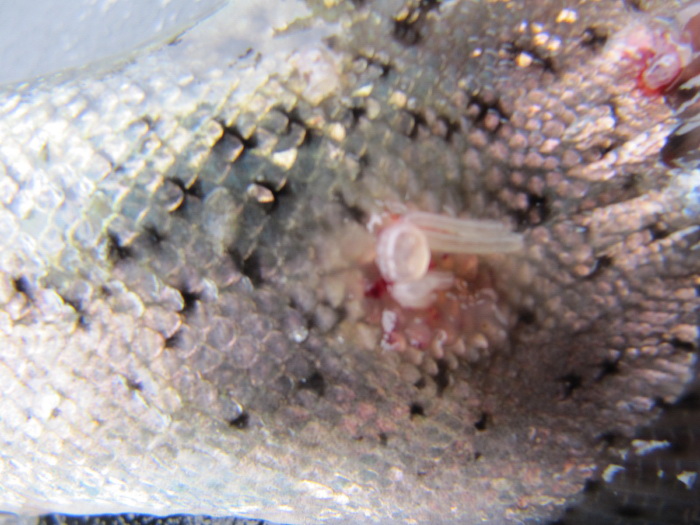
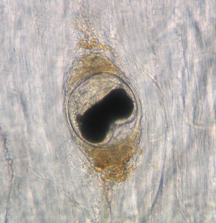
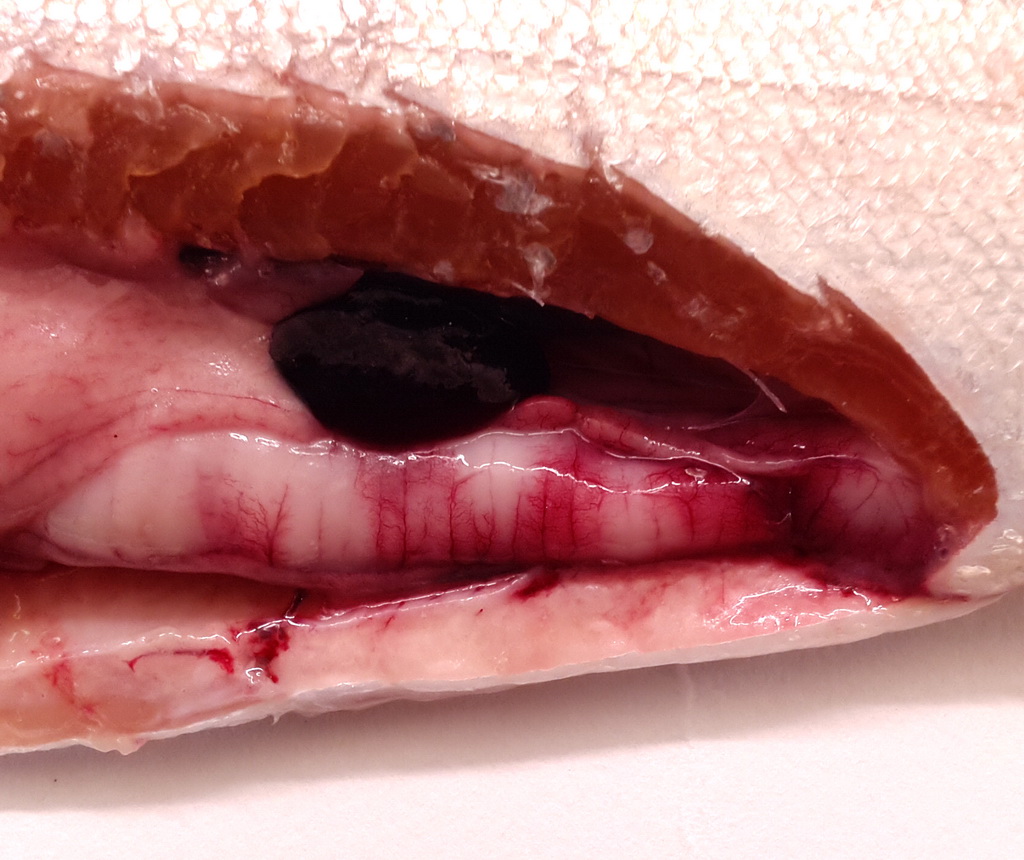
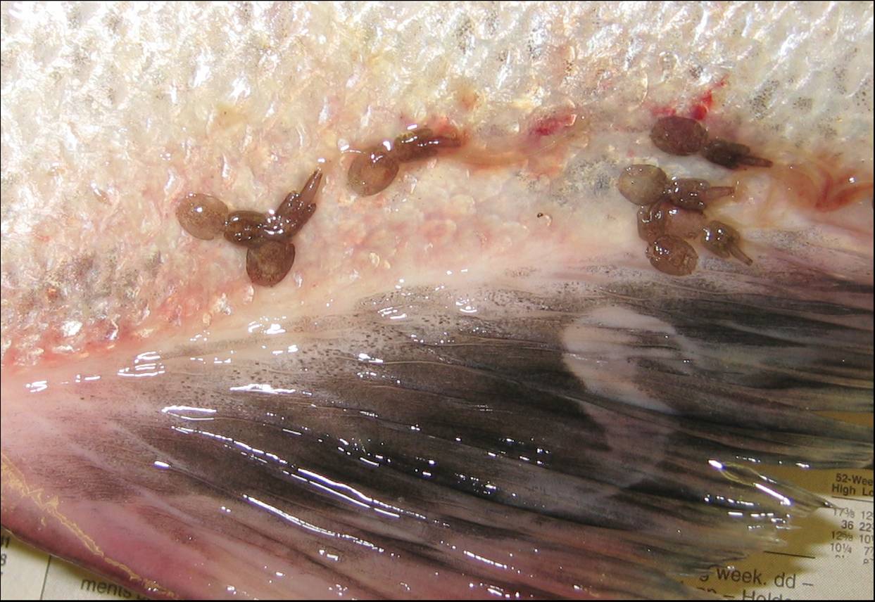
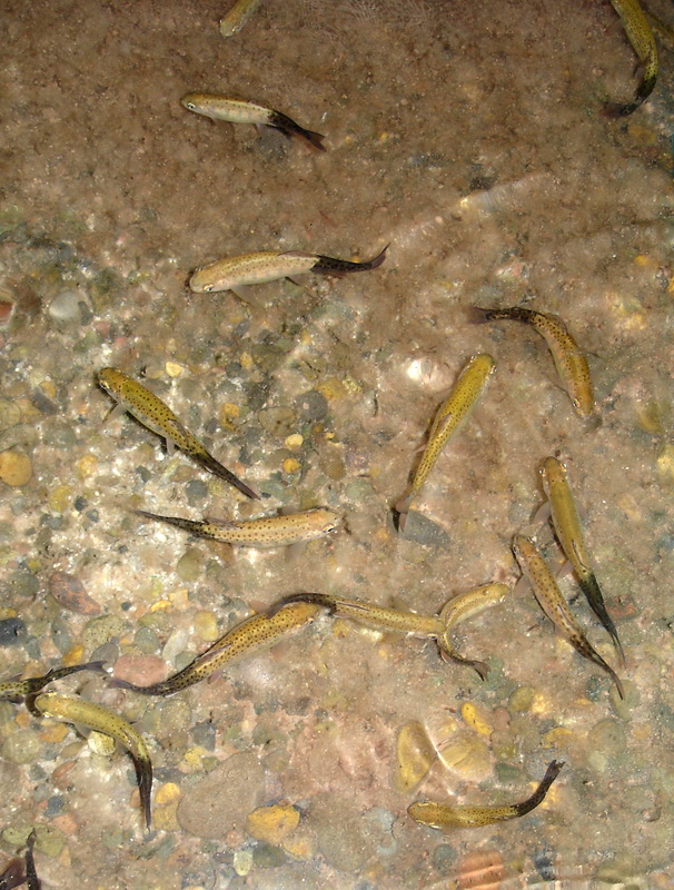
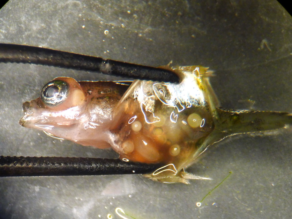
These are all of the pathogen images in our database. Click a pathogen name to jump to the pathogen info page. Click an image to enlarge it, then use the left and right arrows to browse.
Bothriocephalus sp.…
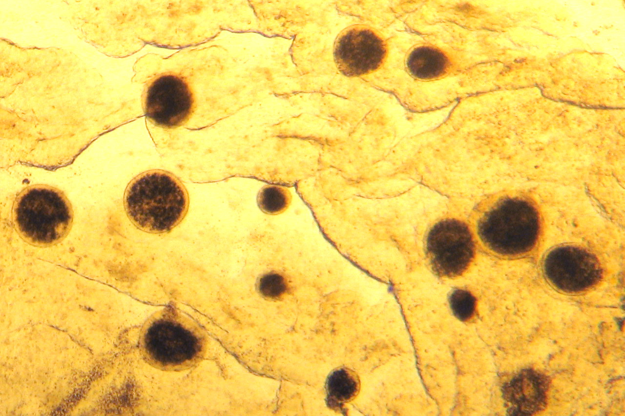
Myxobolus insidiosu…
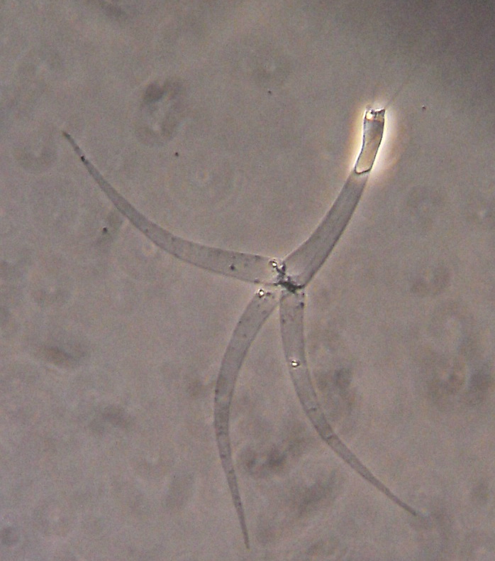
Echinorhynch…
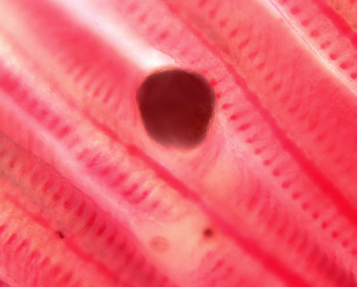
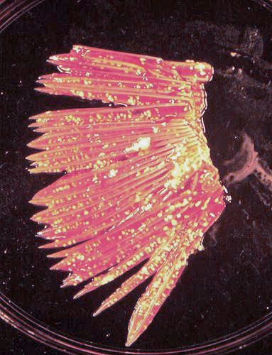
Myxobolus cerebrali…
Flav…
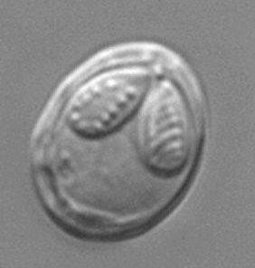
Myxobolus cerebrali…
…
…
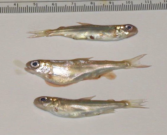
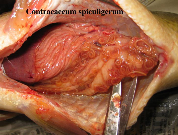
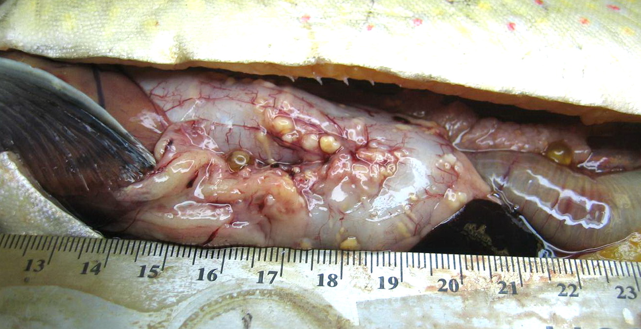
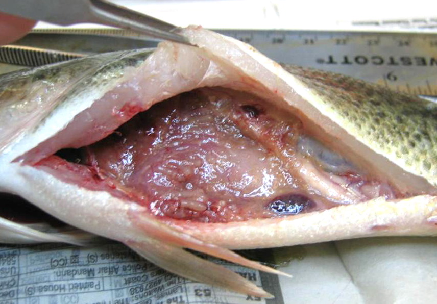
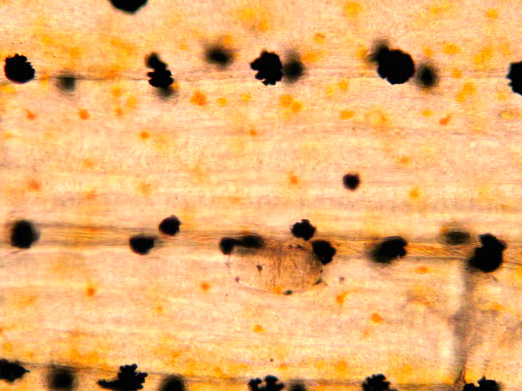
Contracaecum
Diphyllob…
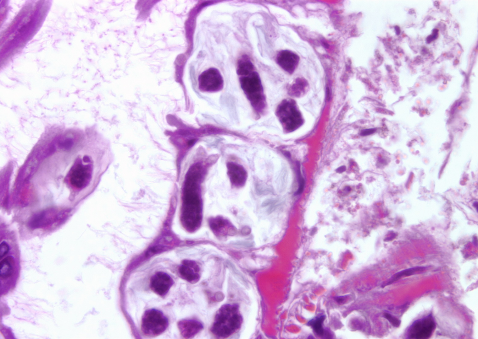
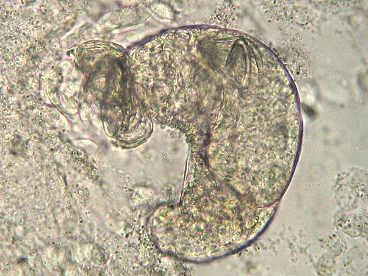
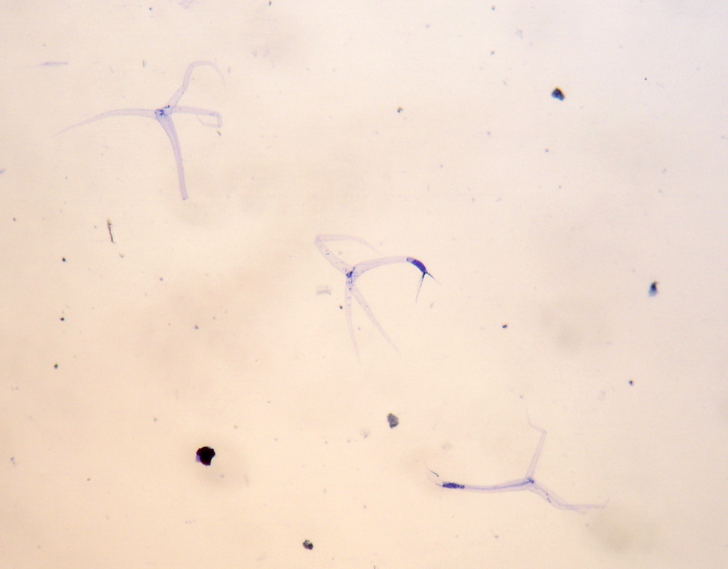
Copepods
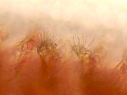
Trichophrya
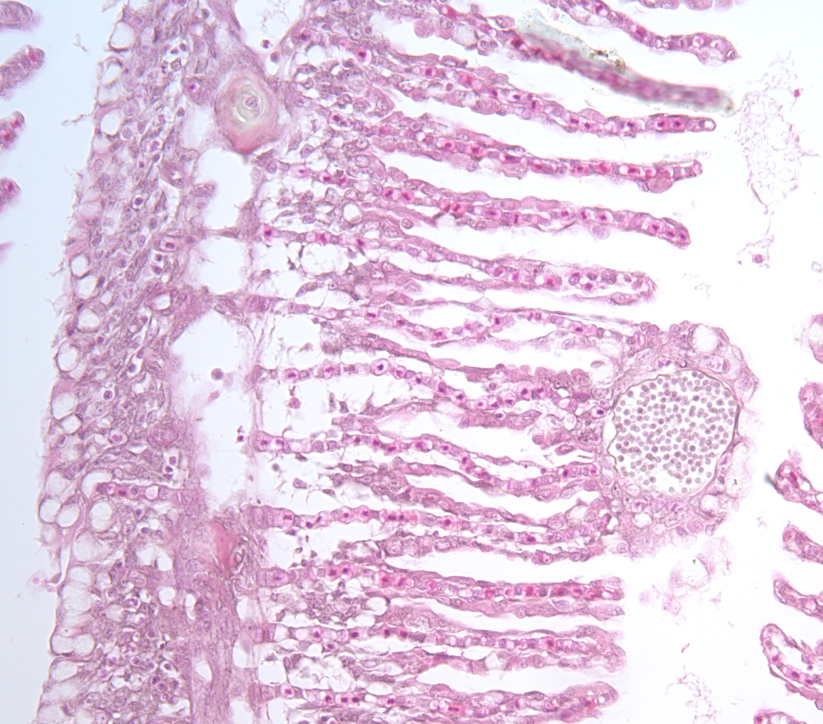
Myxobolus cerebrali…
Copepods
Nanophyetus
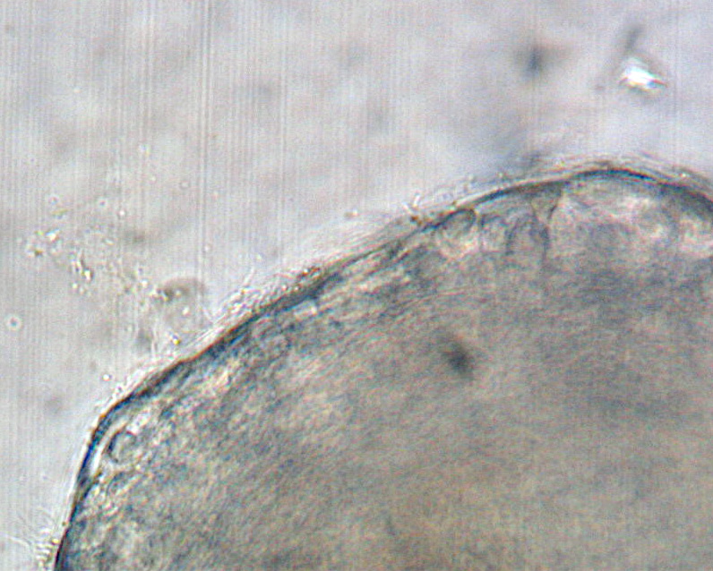
Ceratonova shasta
Copepods
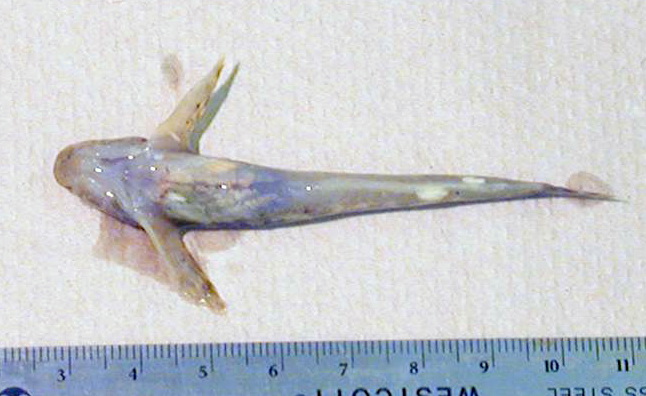
Ichthyobodo
Myxobolus cerebrali…
Ambiphrya
Leeches (Hirudineans…
Henneguya salminic…
Koi Herpesvirus (KHV…
Ichthyo…
Loma
Myxobolus sp.
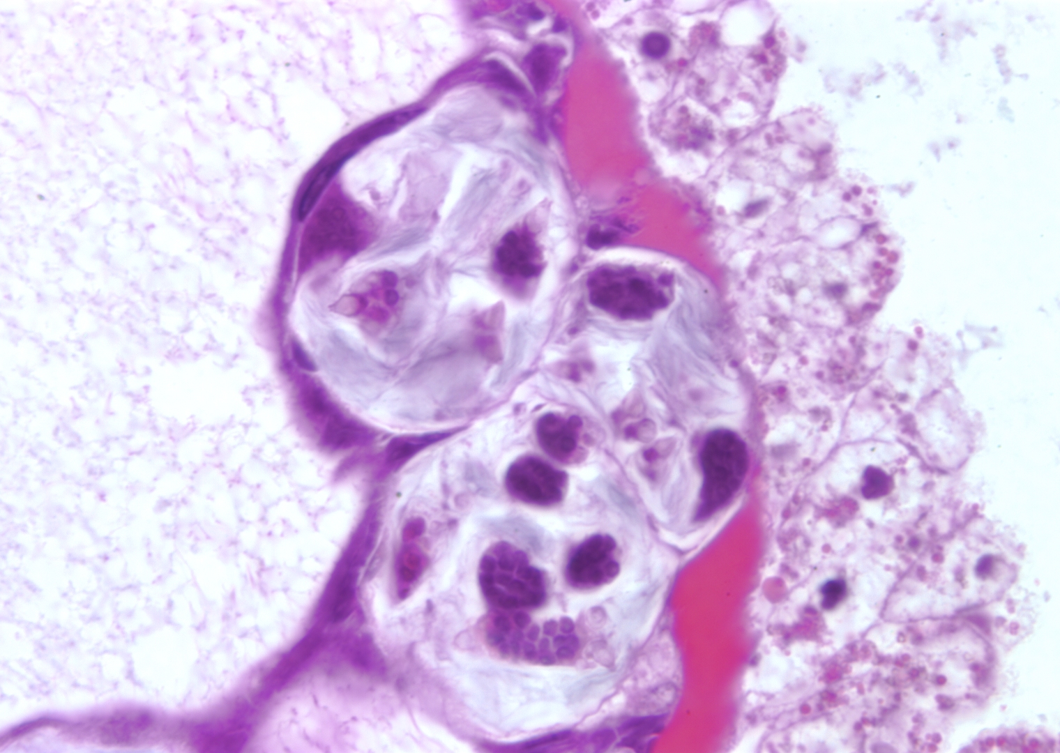
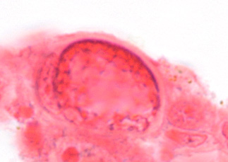
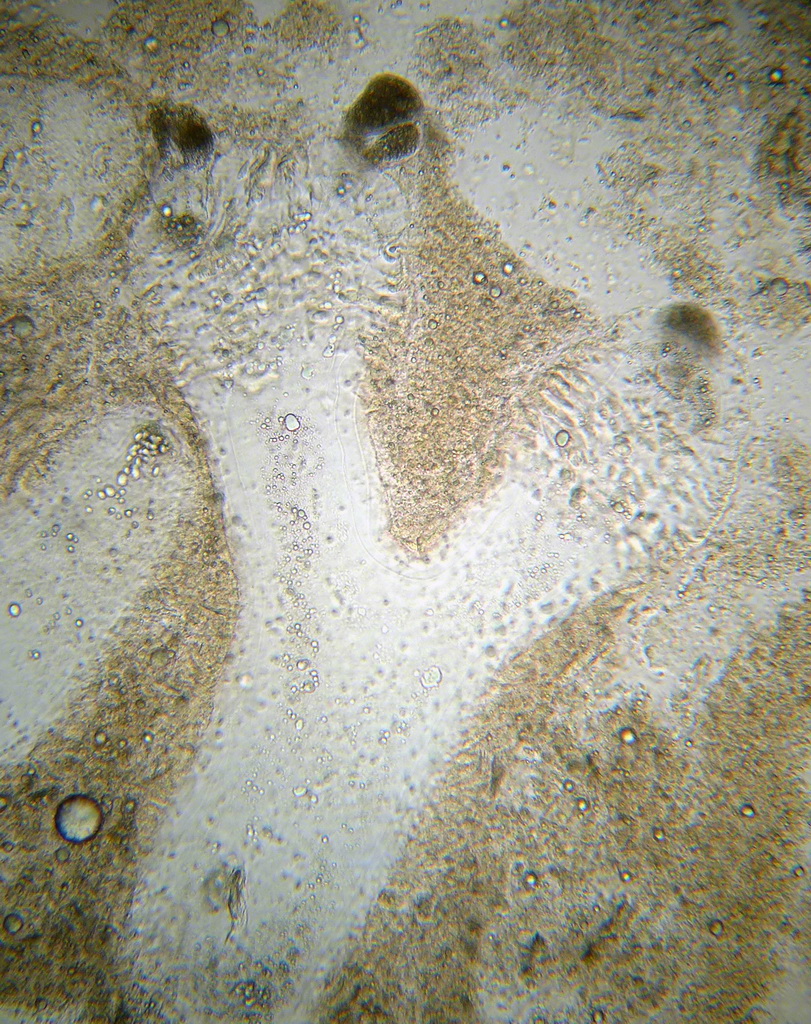
Dermocystidium
Trypanosomes
Rhadinorhynchus sp.…
Proteocephalus
Posthodiplo…
Crepidostomum
Myxobolus cerebrali…
Gyrodactylus
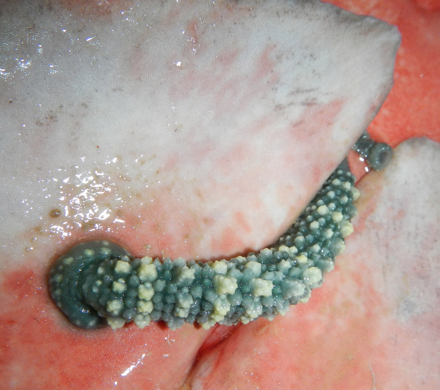
Dermocystidium
Myxobolus cerebrali…
Myxobolus squamalis…
Ichthyo…
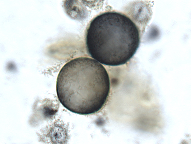
Glugea
…
Neascus
…
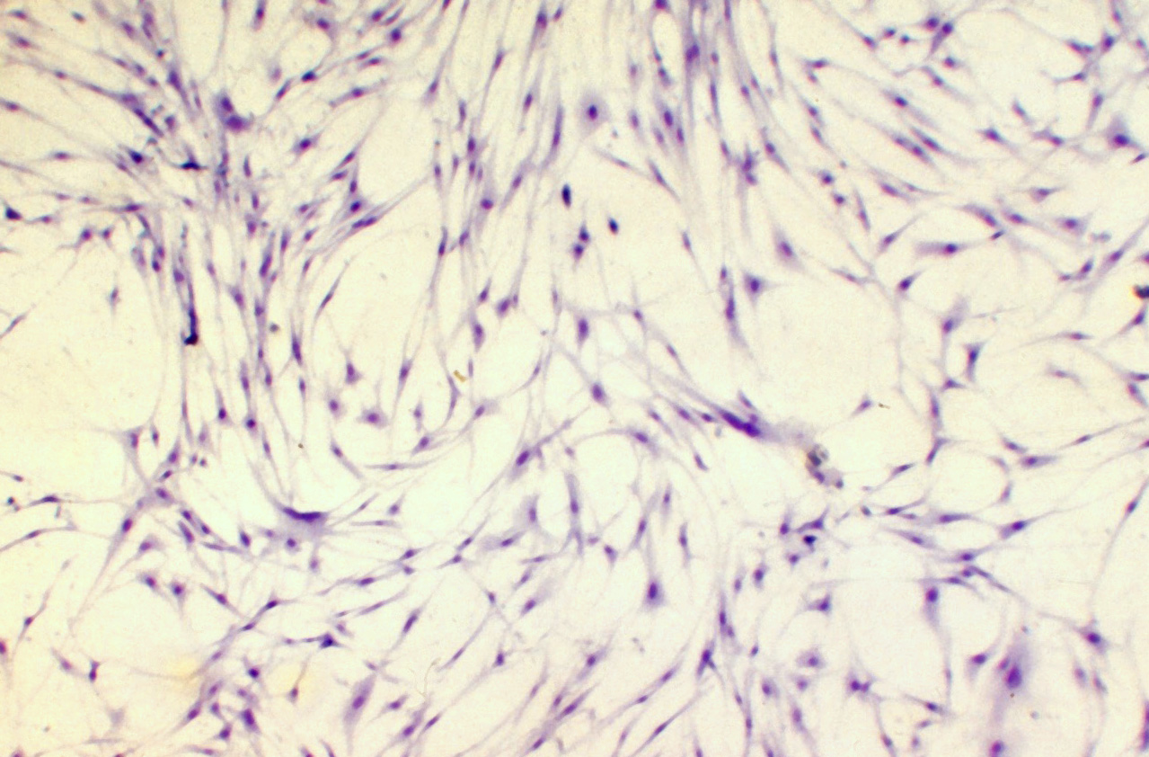
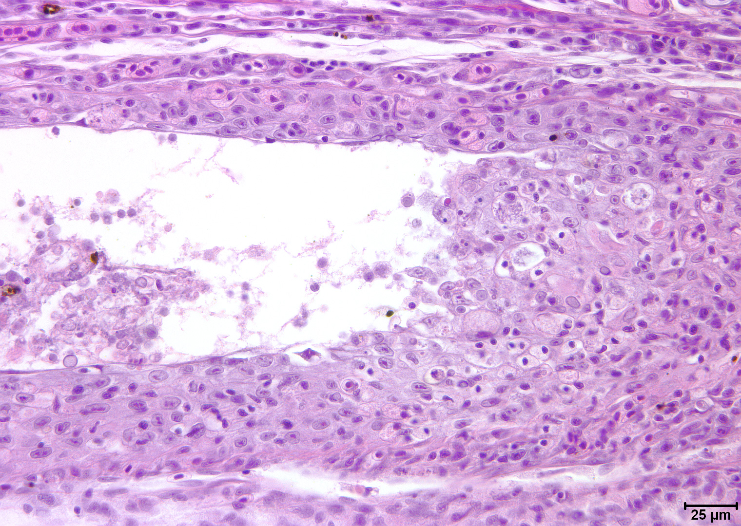
Ichthyophonus
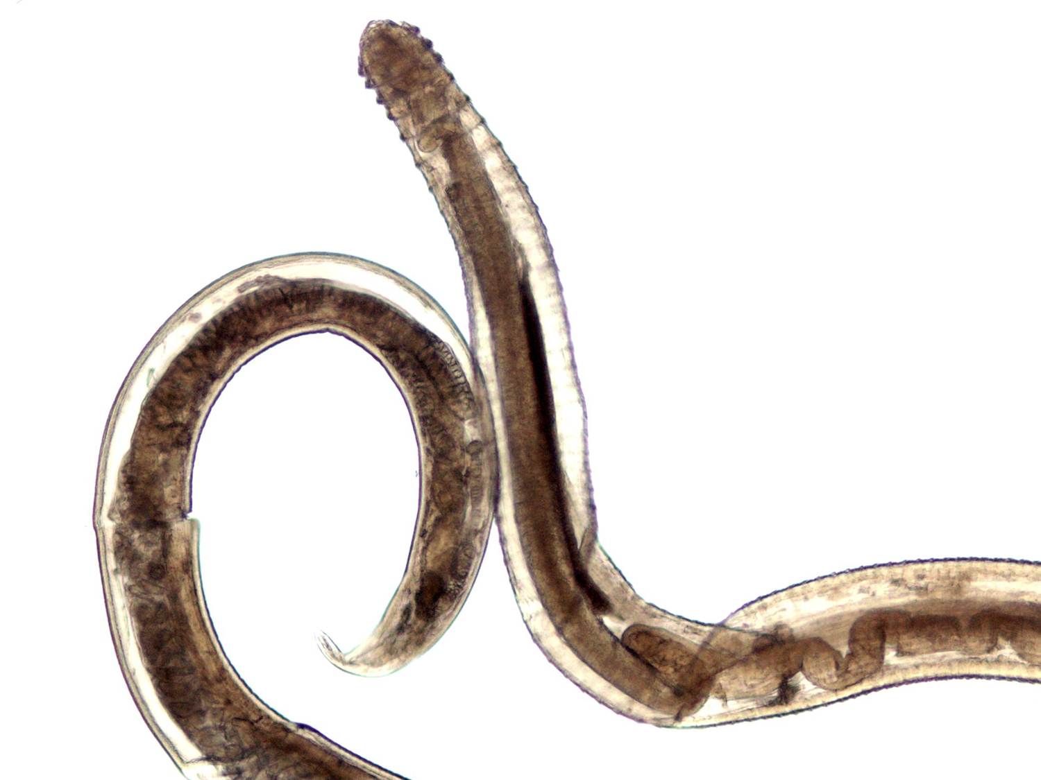
Spinitectus
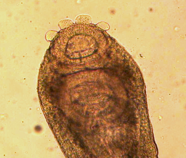
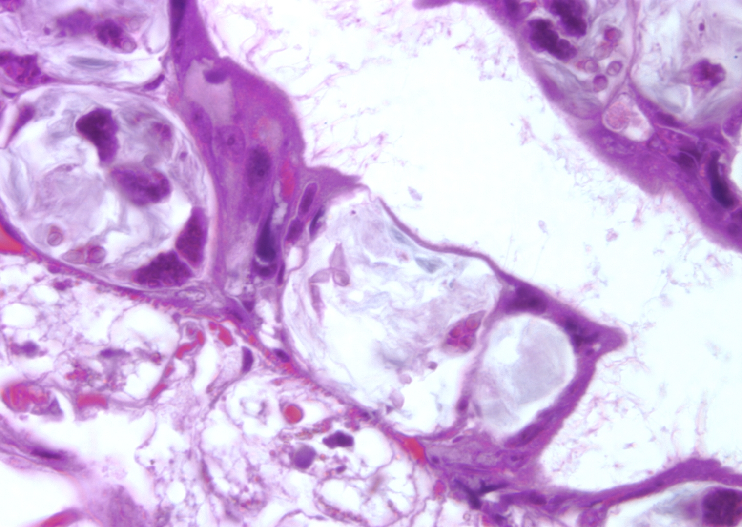
Trematodes (fluk…
Myxobolus cerebrali…
Myxobolus cerebrali…
Nanophyetus
Ceratonova shasta
Trematodes (fluk…
Pagination
Previous page
8
Next page
RSS feed