Skip to main content
Fish Pathogens
Menu
Search
Search
Main navigation
Image Galleries
Pathogens
Pathogens sub-navigation
10 Common Pathogens
What's That Spot?
Ask an Expert
FAQ
Search
Search
Search
User account menu
Log in
Primary tabs
Common Pathogens
All Pathogens
Hosts
Field Sites
Videos
Contributed
All Pathogens
These are all of the pathogen images in our database. Click a pathogen name to jump to the pathogen info page. Click an image to enlarge it, then use the left and right arrows to browse.
Salvelinema walkeri…
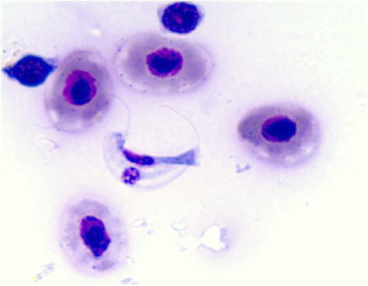
Ichthyobodo
Ichthyo…
Ceratonova shasta
…
Echinorhynch…
Apiosoma
Trichophrya
Bothriocephalus sp.…
Aeromonas salmonic…
Henneguya salminic…
Ambiphrya
Clinostomum (Yel…
non-pa…
Flav…
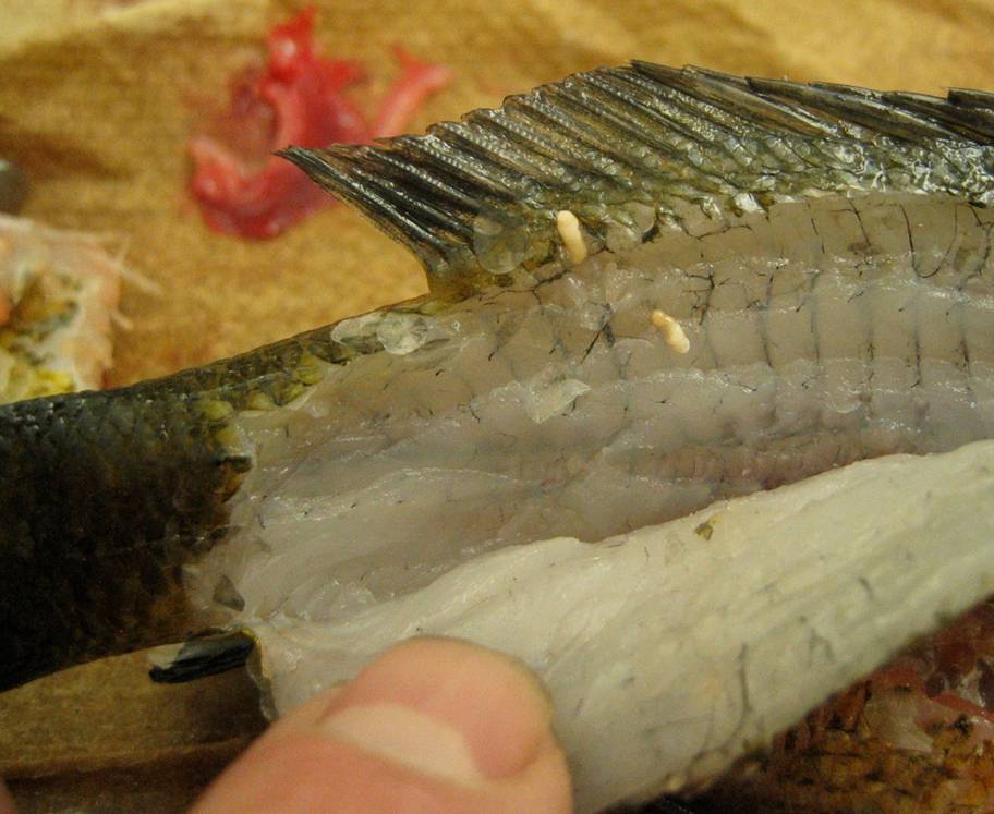
Trematodes (fluk…
Nematodes (round w…
Ceratonova shasta
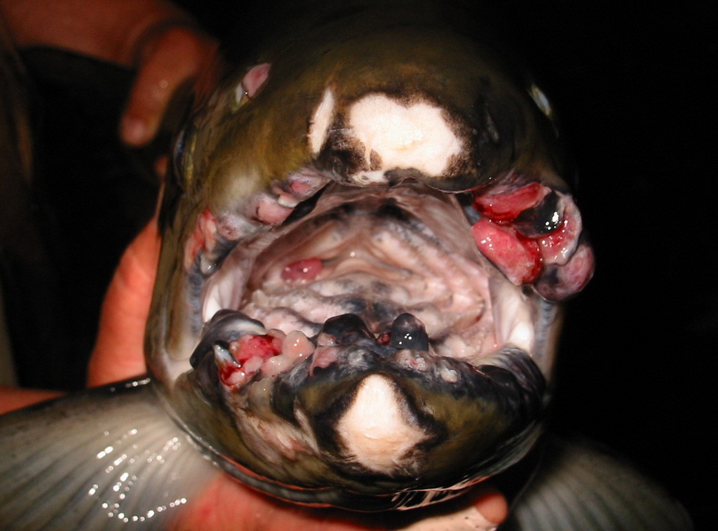
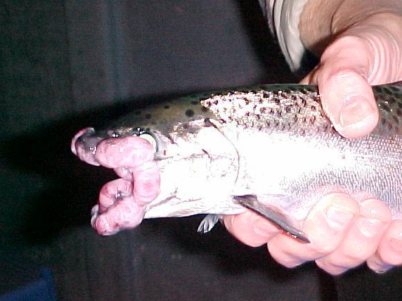
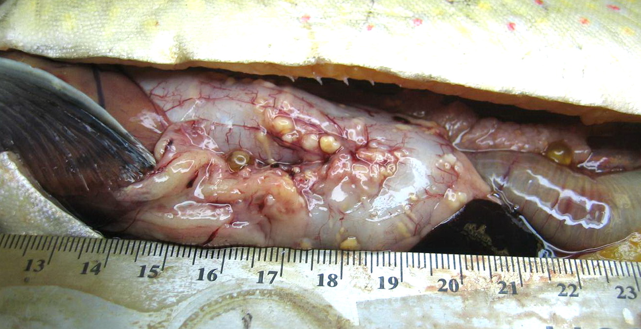
Tumor-like lumps
…
Trematodes (fluk…
Tumor-like lumps
Clinostomum (Yel…
Cryptobia salmosit…
Diphyllob…
Vibrio
Echinochasmus
Flav…
Trematodes (fluk…
Ichthyobodo
Ceratonova shasta
Trematodes (fluk…
Fungi and fun…
Myxobolus cerebrali…
White Sturgeon…
Salmincola
Bacteria
…
Neascus
Clinostomum (Yel…
non-pa…
Trematodes (fluk…
Trematodes (fluk…
Gloch…
Neascus
Ichthyobodo
Ichthyophonus
Trematodes (fluk…
Trematodes (fluk…
…
Pagination
1
Next page
RSS feed