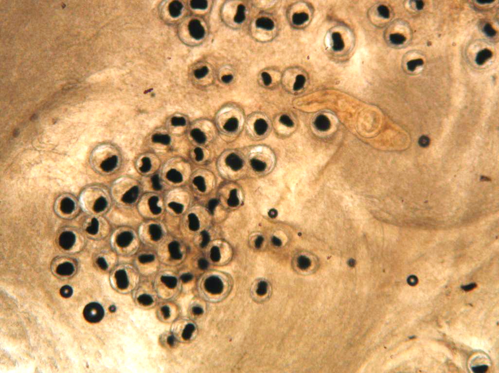
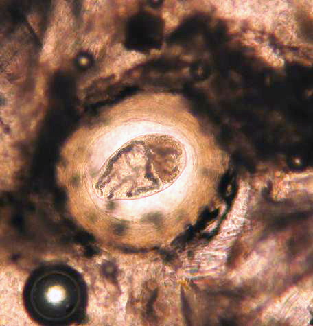
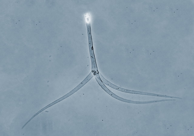
| nanophyetus_salmonicola_CB06.jpg | All Pathogens, Fact Sheet Images | |
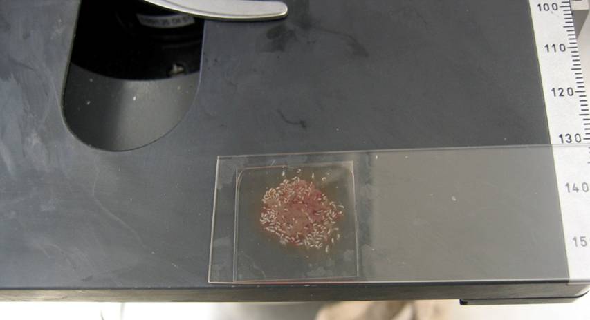
 | Renibacterium_3.jpg | All Pathogens | |
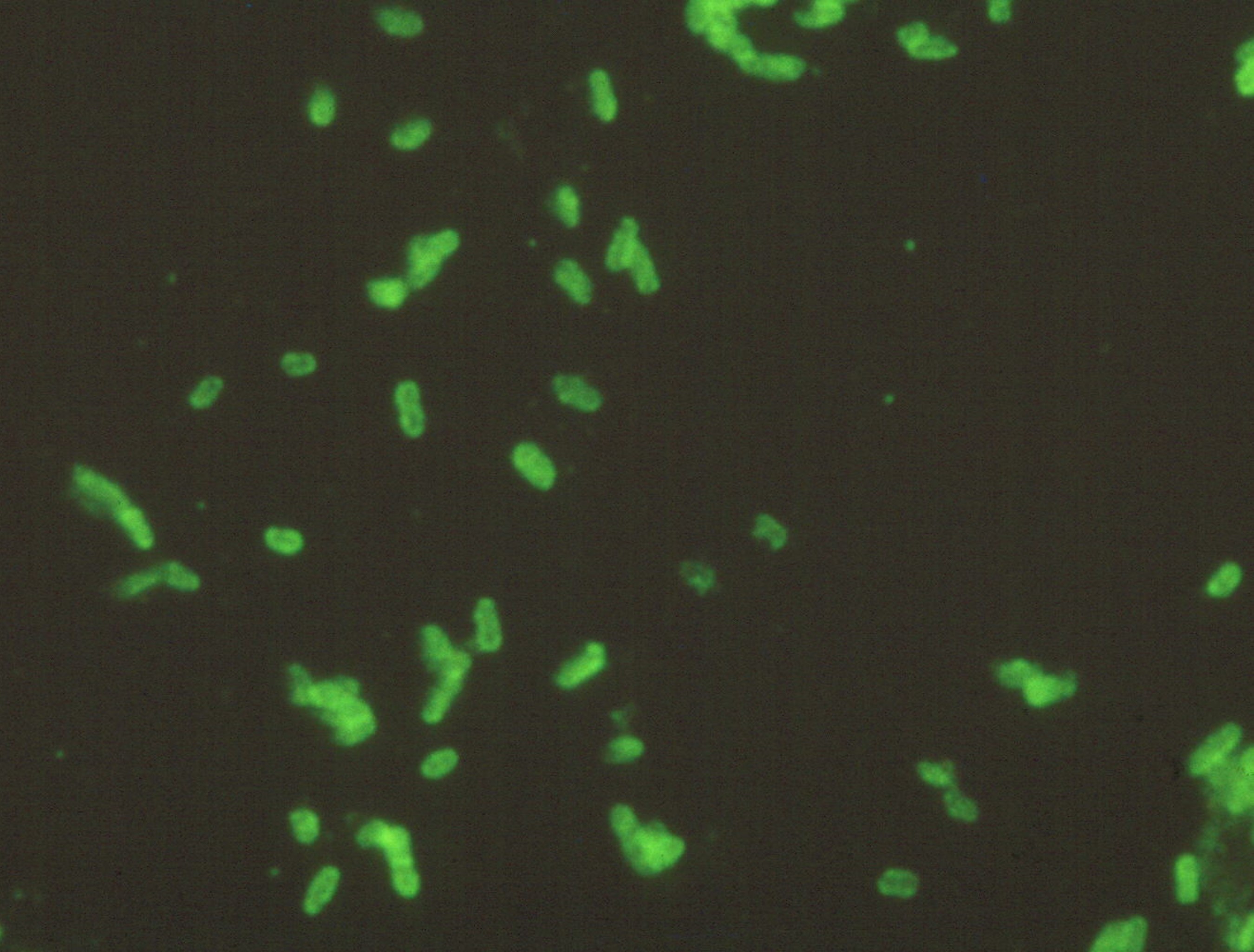
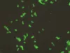 | Renibacterium_1.jpg | All Pathogens | |
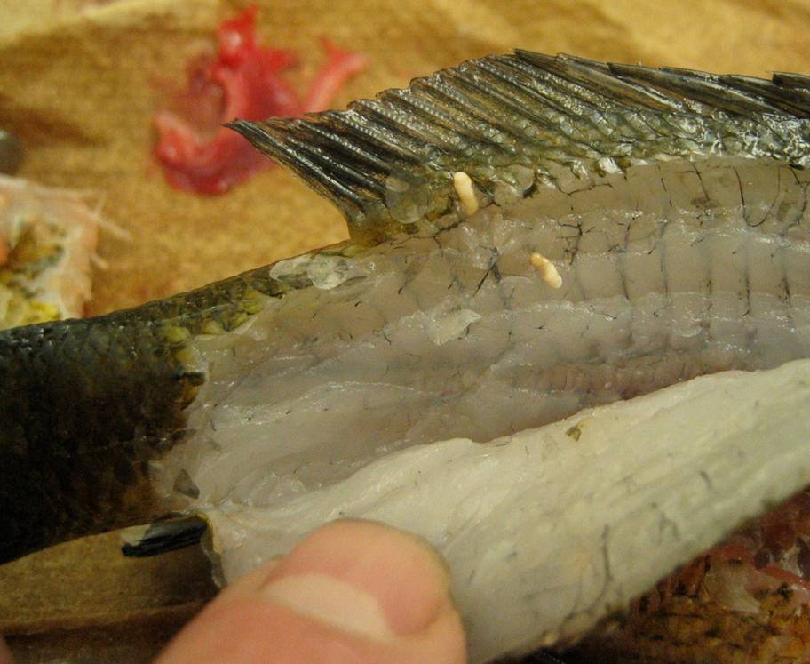
 | Clinostomum_05.jpg | All Pathogens, Fact Sheet Images | |
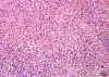 | Slide # 40 Necrotizing steatitis Flavobacterium psychrophilus H&E 400X.jpg | All Pathogens, Fact Sheet Images | |
 | nanophyetus_salmonicola_CB07.jpg | All Pathogens, Fact Sheet Images | |
 | Clinostomum_02.jpg | All Pathogens, Fact Sheet Images | |
 | nanophyetus_salmonicola_CB08.jpg | All Pathogens, Fact Sheet Images | |
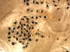
 | BO_Ichthyophonus_x20_37.jpg | All Pathogens | |
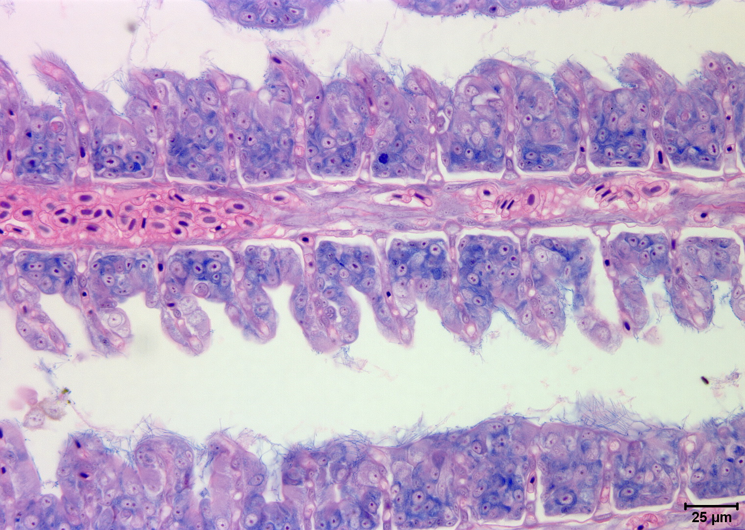
 | Slide # 37 Gills Flavobacterium branchiophilus Giemsa 400X.jpg | All Pathogens | |
| | nanophyetus_salmonicola_CB08.jpg | All Pathogens, Fact Sheet Images | |
 | Clinostomum_04.jpg | All Pathogens, Fact Sheet Images | |
 | Slide # 36 Gills Flavobacterium branchiophilus H&E 400X.jpg | All Pathogens | |
 | BO_Ichthyophonus_x20_38.jpg | All Pathogens | |
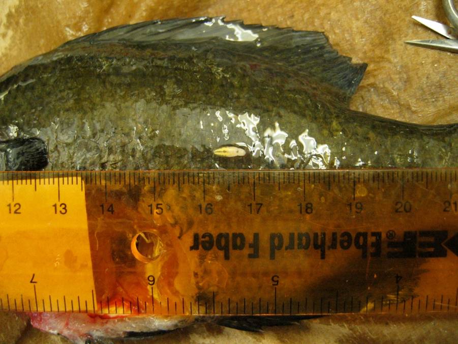
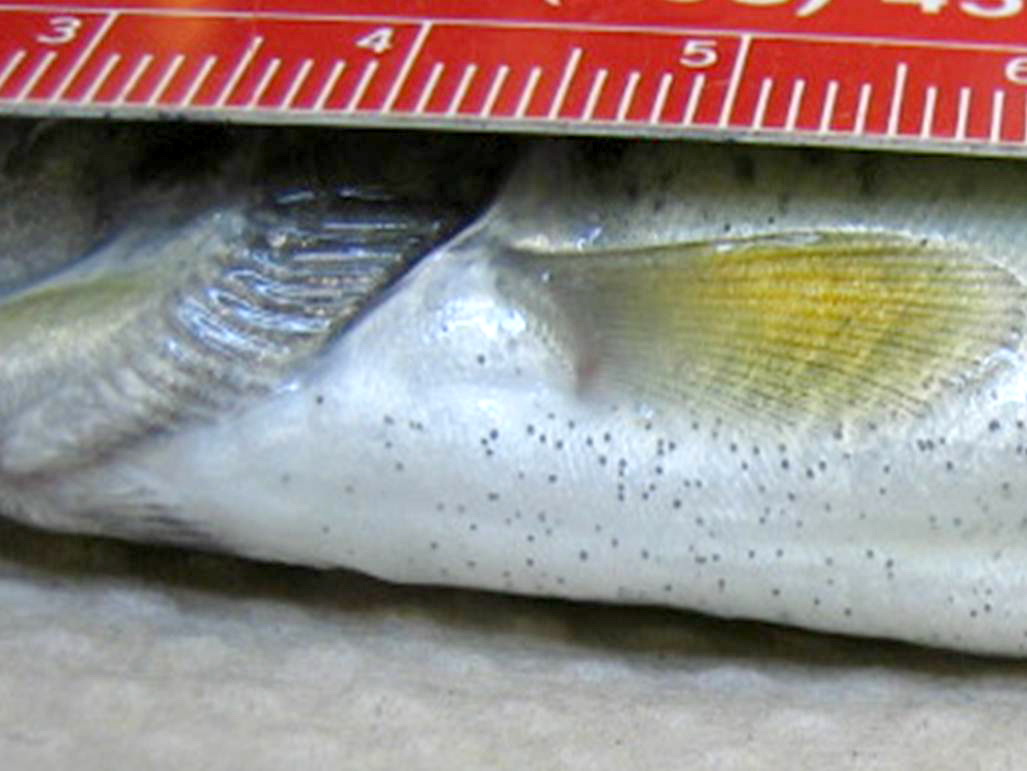
 | nanophyetus_salmonicola_CB09.jpg | All Pathogens, Top 10, Fact Sheet Images | yellow perch |
 | Slide # 36 Gills Flavobacterium branchiophilus H&E 400X 2.jpg | All Pathogens | |
 | nanophyetus_salmonicola_CB12.jpg | All Pathogens, Fact Sheet Images | |
 | BO_Loma_x63_19.jpg | All Pathogens | |
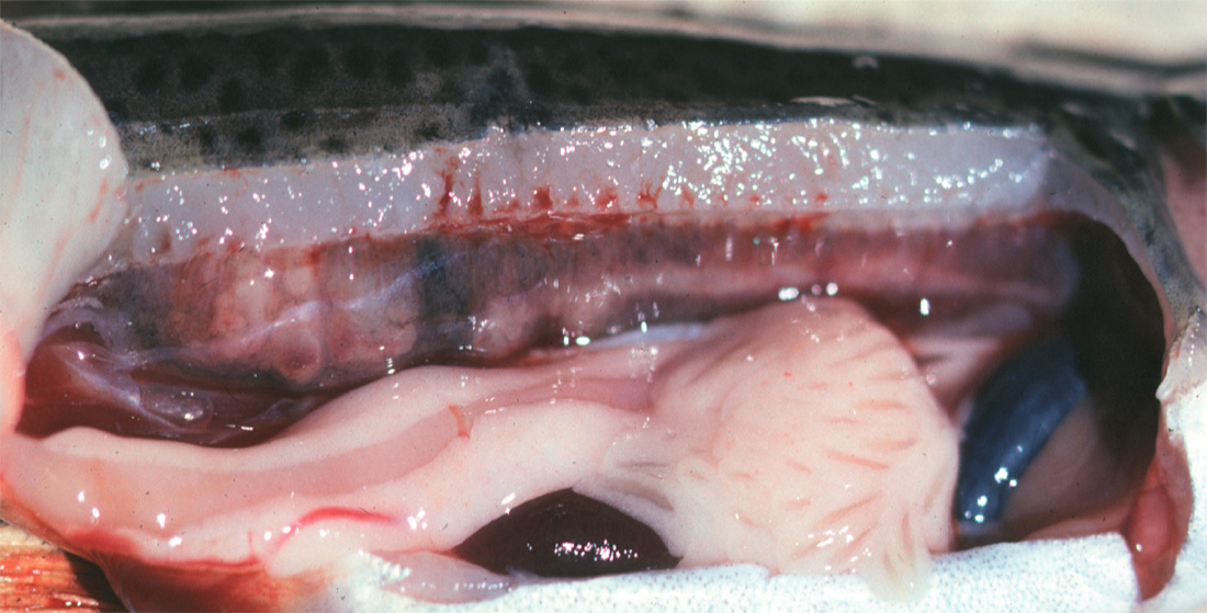
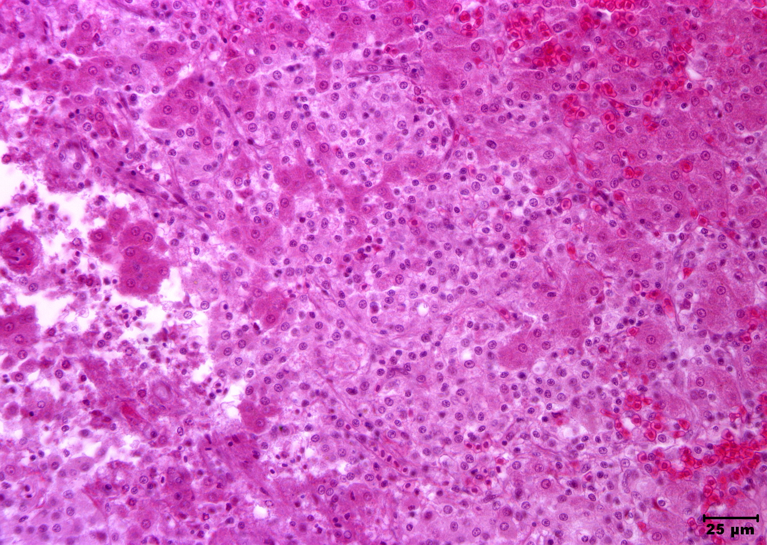
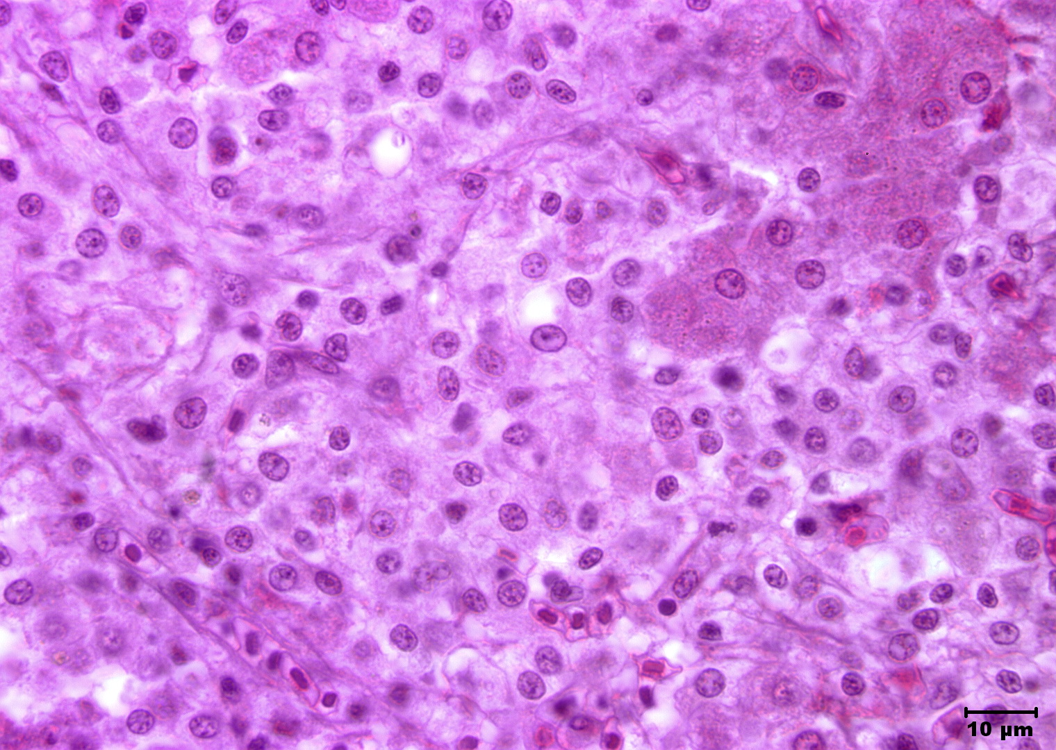
 | Slide # 33 Liver Hepatocellular Necrosis Edwardsiella ictaluri H&E 400X.jpg | All Pathogens, Fact Sheet Images | |
 | Clinostomum_03.jpg | All Pathogens, Top 10, Fact Sheet Images | |
 | nanophyetus_salmonicola_CB11.jpg | All Pathogens, Fact Sheet Images | |
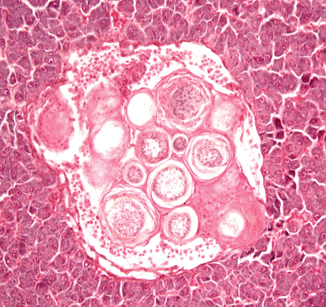
 | neascus_CB01.jpg | All Pathogens, Fact Sheet Images | |
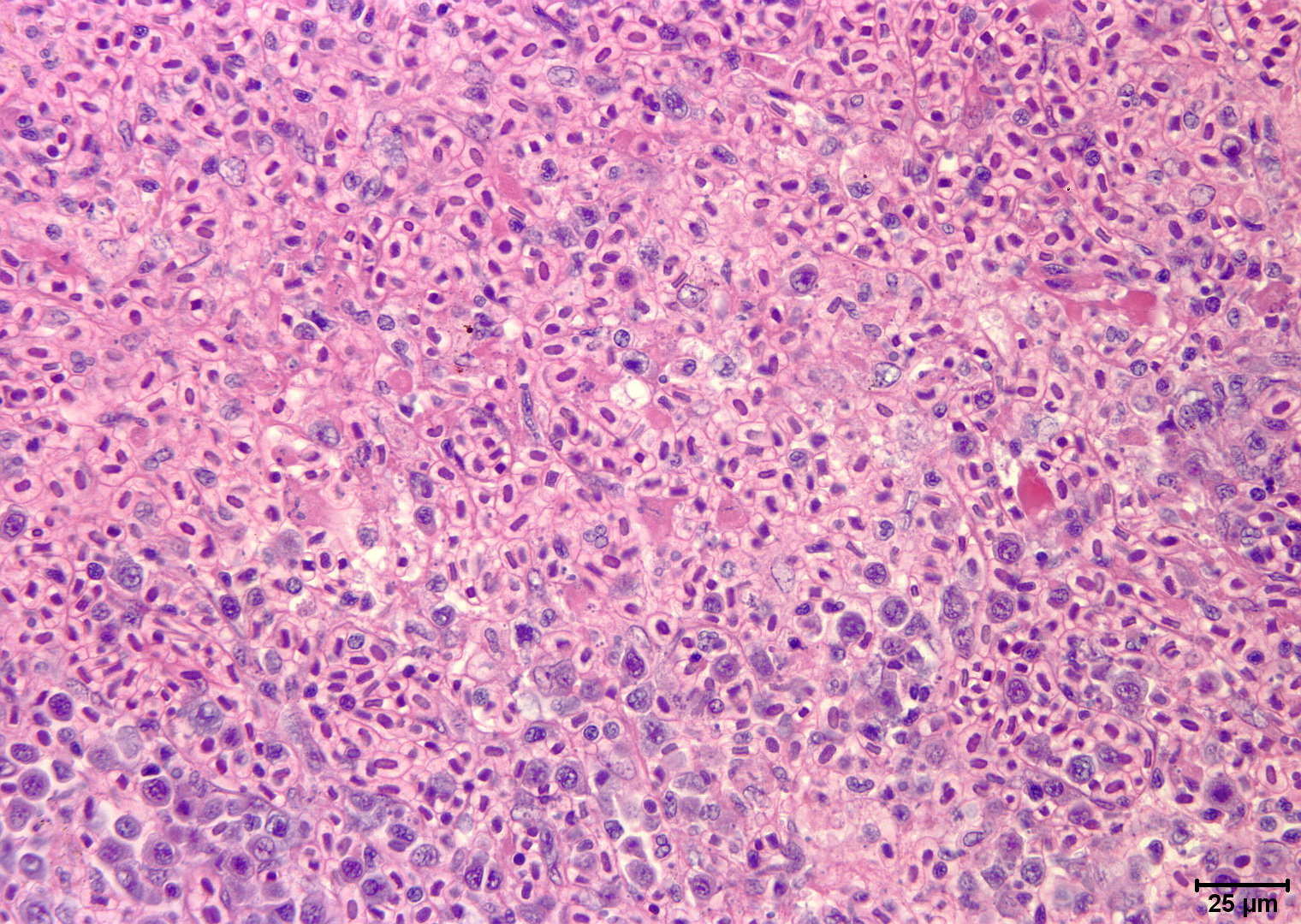
 | Slide # 33 Liver Hepatocellular Necrosis Edwardsiella ictaluri H&E Oil 1000X.jpg | All Pathogens, Fact Sheet Images | |
 | nanophyetus_salmonicola_CB10.jpg | All Pathogens, Fact Sheet Images | |
 | Slide # 19 Kidney Aeromonas salmonicida Giemsa 400X.jpg | All Pathogens | |
 | BO_Loma_x63_18.jpg | All Pathogens | |
 | fishpathogens_.jpg | All Pathogens, Contributed Images | rainbow trout |
 | DigsFish_Zeylandicobdella-est-cod.jpg | All Pathogens, Contributed Images | estuary cod - Epinephelus coioides |
 | DigsFish_29-nov-12-Pontobdella-loricata.jpg | All Pathogens, Contributed Images | shark - Carcharhinus sp. |
 | SA_Generic_TAM.jpg | All Pathogens, Fact Sheet Images | |