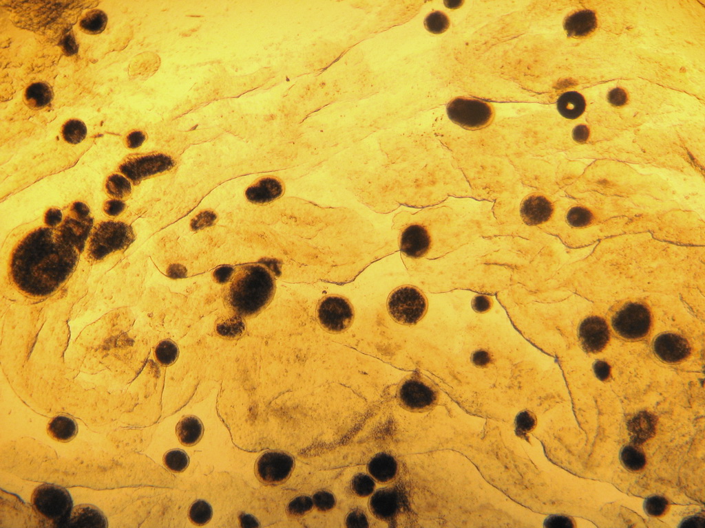
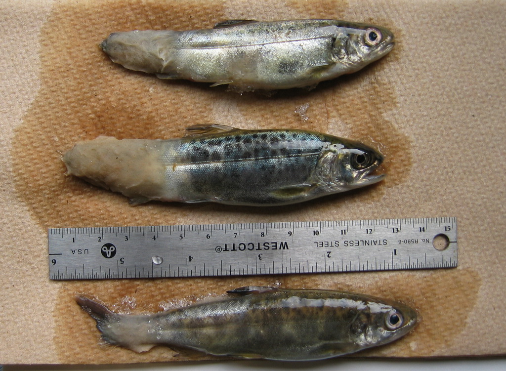
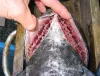
 | CB_CoBLkSpt30.jpg | All Pathogens, Fact Sheet Images | coho salmon |
 | CB_CohoBlk_spt4.jpg | All Pathogens, Fact Sheet Images | coho salmon |
 | CB_Gtrem17.jpg | All Pathogens, Fact Sheet Images | |
 | CB_HeavyIch2.jpg | All Pathogens, Fact Sheet Images | |
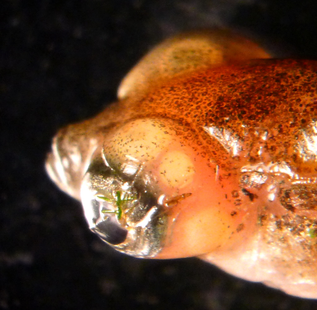
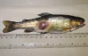
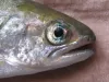 | CB_Blue_eye_3.jpg | All Pathogens, Fact Sheet Images | rainbow trout |
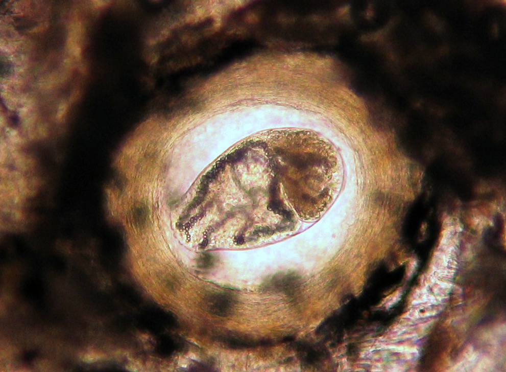
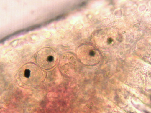
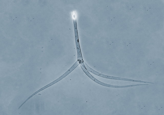
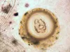
 | P1060629_diplostomum.jpg | All Pathogens, Fact Sheet Images | threespine stickleback |
 | CB_Sanguin8.jpg | All Pathogens, Fact Sheet Images | |
 | CB_HeavyIch.jpg | All Pathogens, Fact Sheet Images | |
 | CB_PGE_Fung1.jpg | All Pathogens, Fact Sheet Images | |
 | CB_Cop_adult_gill.jpg | All Pathogens, Fact Sheet Images | |
 | CB_BLK_SPT1.jpg | All Pathogens, Fact Sheet Images | rainbow trout |
 | CB_CRYTO75.jpg | All Pathogens, Fact Sheet Images | |
 | P1060631_diplostomum.jpg | All Pathogens, Fact Sheet Images | threespine stickleback |
| | CB_Blue_eye_3.jpg | All Pathogens, Fact Sheet Images | rainbow trout |
 | CB_Kla_ModCWD12.jpg | All Pathogens, Fact Sheet Images | |
 | SA_Generic_TAM.jpg | All Pathogens, Fact Sheet Images | |
 | Renibacterium_2.png | All Pathogens, Fact Sheet Images | |
 | BO_Dermocystidium_x63_25.jpg | All Pathogens, Fact Sheet Images | |
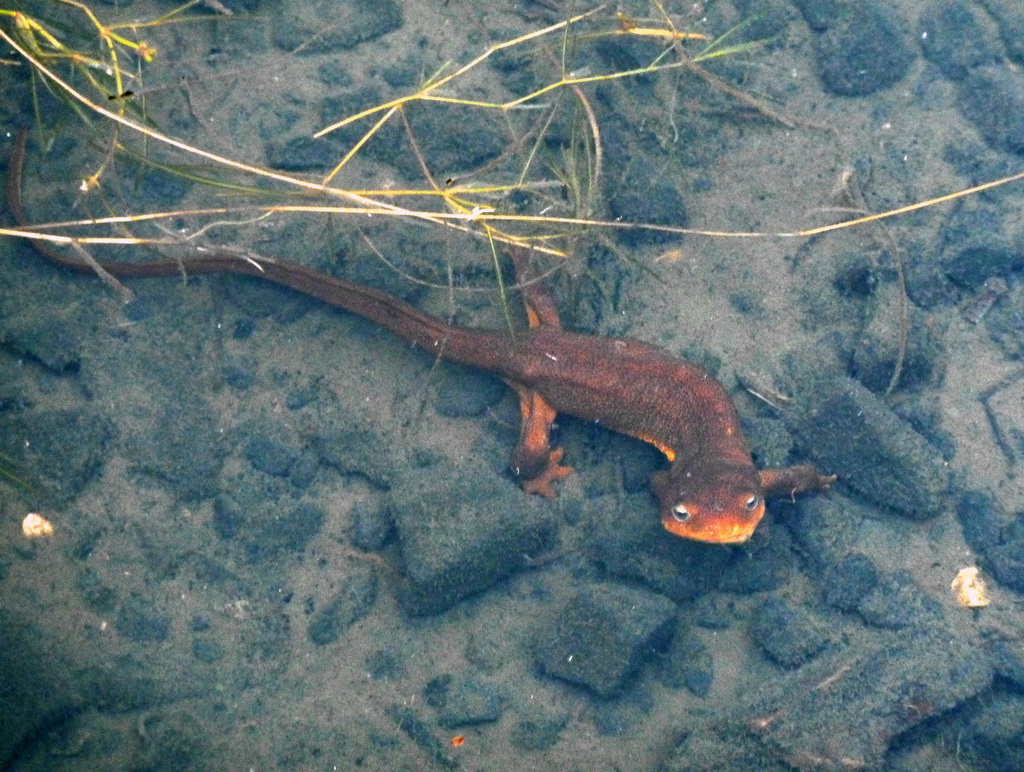
 | P1000836_newt.jpg | Hosts and other organisms, Fact Sheet Images | rough-skinned newt; salamander |
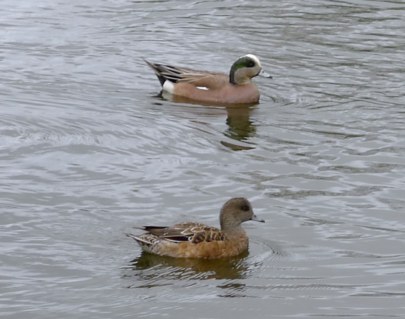
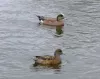 | P1080719_widgeon.jpg | Hosts and other organisms, Fact Sheet Images | American wigeon |
 | CB_IMG_5023.jpg | All Pathogens, Fact Sheet Images | |
 | CB_Apiostoma_siletz_Good3.jpg | All Pathogens, Fact Sheet Images | |
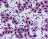
 | CB_Trich7.jpg | All Pathogens, Fact Sheet Images | |
 | CB_146_4665.jpg | All Pathogens, Fact Sheet Images | threespine stickleback |
 | RH_chin_fcolumn.jpg | All Pathogens, Fact Sheet Images | Chinook salmon |
 | CB_Glochidia.jpg | All Pathogens, Fact Sheet Images | |
 | CB_strawberry.jpg | All Pathogens, Fact Sheet Images | rainbow trout |
 | CB_IMG_5012.jpg | All Pathogens, Fact Sheet Images | |
 | CB_Gill_amoeb33.jpg | All Pathogens, Fact Sheet Images | |
 | CB_IMG_5021.jpg | All Pathogens, Fact Sheet Images | |