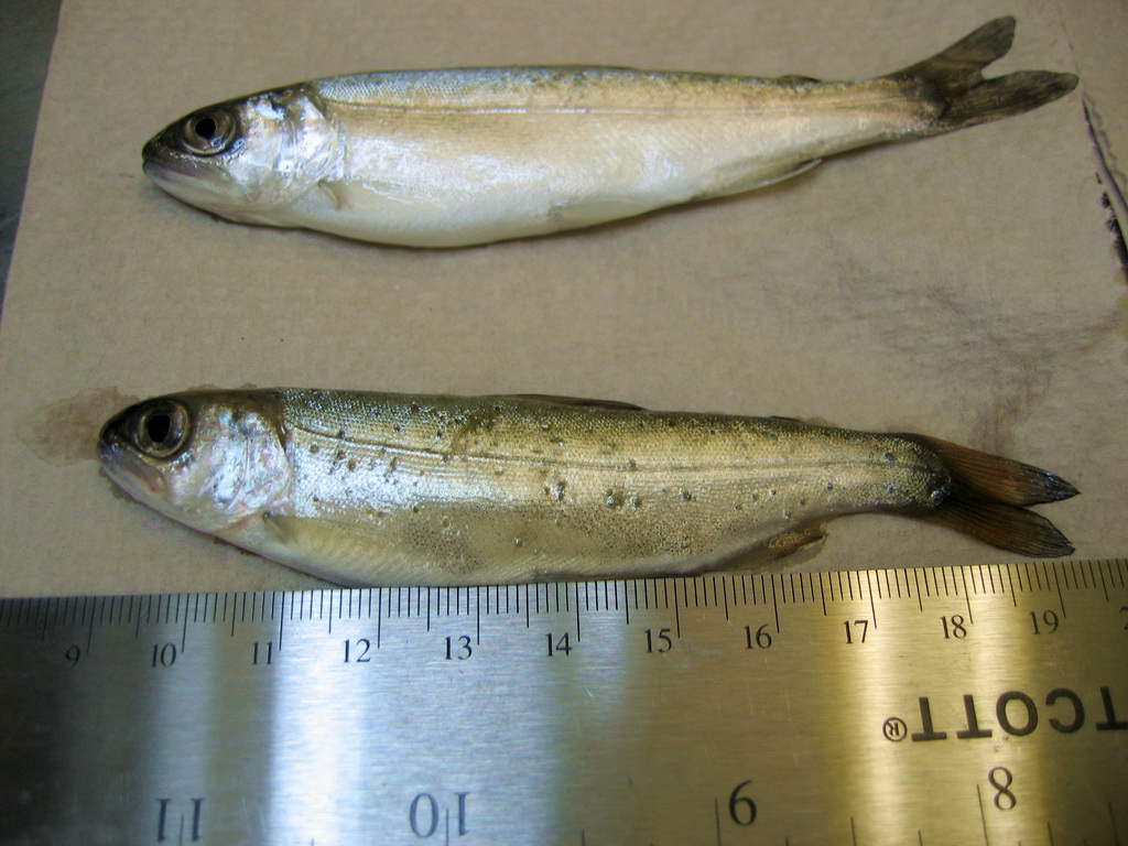
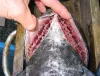
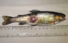
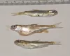
| CB_CohoBlk_spt4.jpg | All Pathogens, Fact Sheet Images | coho salmon |
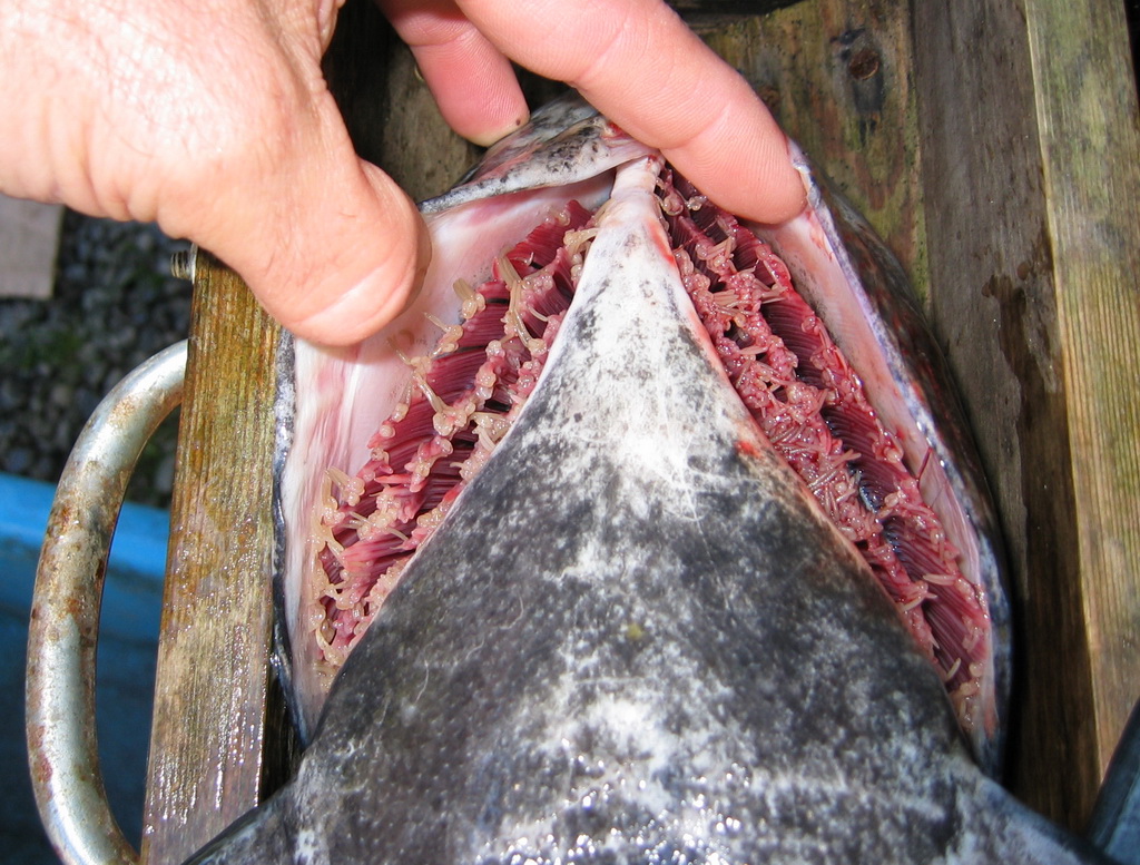
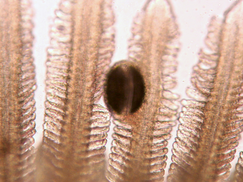
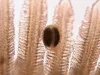
 | CB_Cop_adult_gill.jpg | All Pathogens, Fact Sheet Images | |
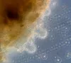
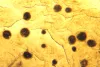
 | CB_CRYTO75.jpg | All Pathogens, Fact Sheet Images | |
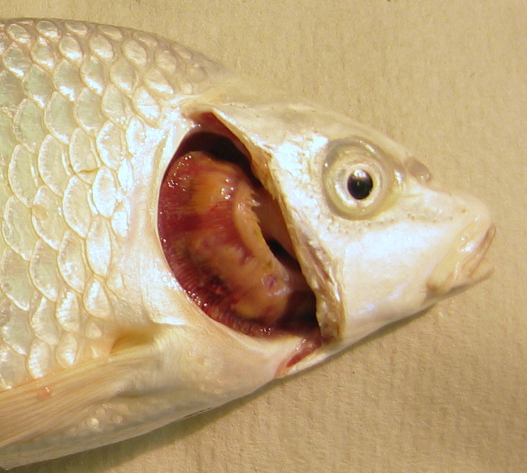
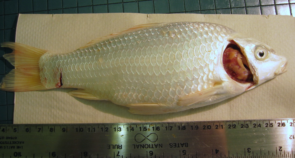
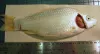
 | CB_GFFR043.jpg | All Pathogens, Fact Sheet Images | goldfish |
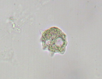
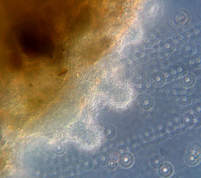
 | CB_Amoeba19.jpg | All Pathogens, Fact Sheet Images | |
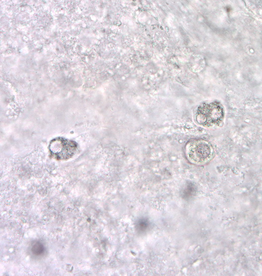
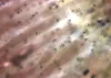
 | CB_Gill_amoeb33.jpg | All Pathogens, Fact Sheet Images | |
 | CB_Glochidia.jpg | All Pathogens, Fact Sheet Images | |
 | CB_FCOLrc05.jpg | All Pathogens, Fact Sheet Images | |
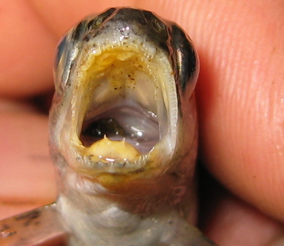
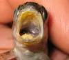 | CB_Mouth_column4.jpg | All Pathogens, Fact Sheet Images | |
 | CB_Koi_Columnaris2b.jpg | All Pathogens, Fact Sheet Images | koi |
 | CB_Koi_Columnaris2a.jpg | All Pathogens, Hosts and other organisms, Fact Sheet Images | koi |
 | CB_Kla_ModCWD12.jpg | All Pathogens, Fact Sheet Images | |
 | CB_HeavyIch.jpg | All Pathogens, Fact Sheet Images | |
 | CB_HeavyIch2.jpg | All Pathogens, Fact Sheet Images | |
 | CB_Gtrem17.jpg | All Pathogens, Fact Sheet Images | |
 | CB_BKD_02_ChS.jpg | All Pathogens, Fact Sheet Images | Spring Chinook salmon |
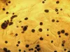
 | Neascus_06.jpg | All Pathogens, Fact Sheet Images | |
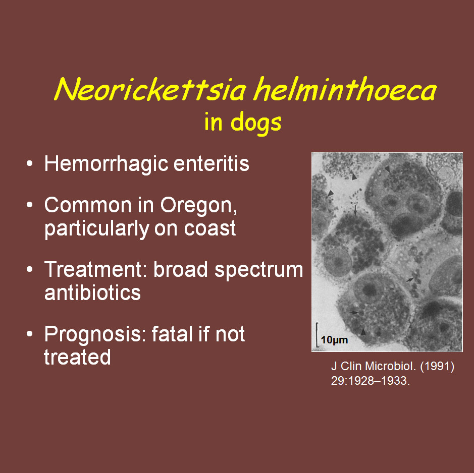
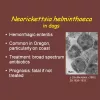 | CB_Neorickettsia_ppt.jpg | Fact Sheet Images, Illustration - organism | Nanophyetus |
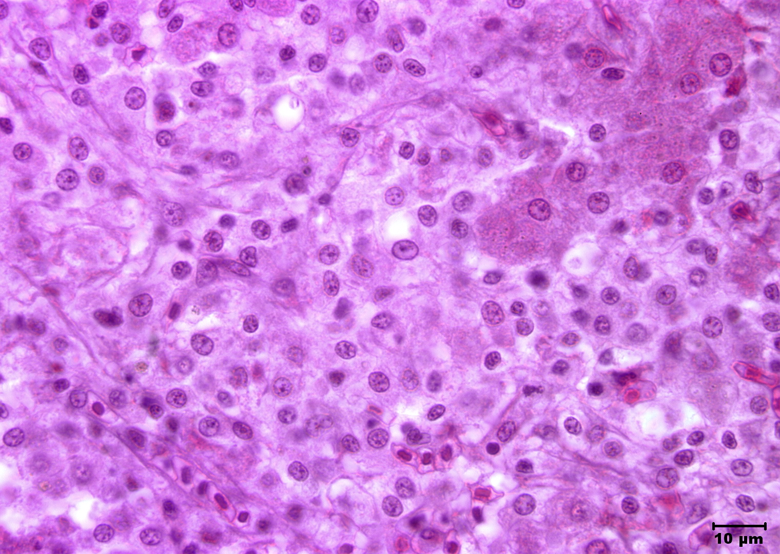
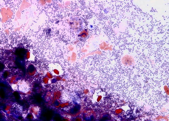
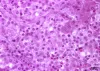 | Slide # 33 Liver Hepatocellular Necrosis Edwardsiella ictaluri H&E Oil 1000X.jpg | All Pathogens, Fact Sheet Images | |
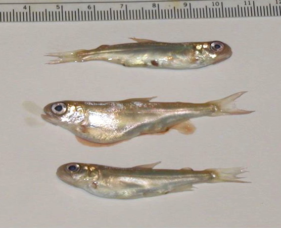
 | CB_Philometroides_01.jpg | All Pathogens, Fact Sheet Images | largescale sucker |
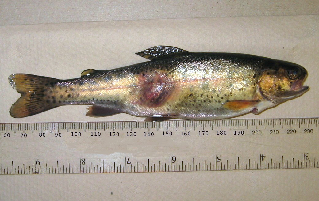
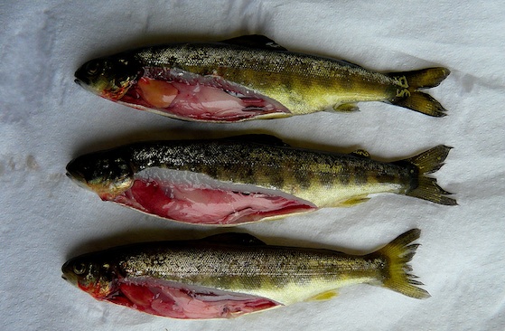
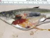
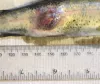 | CB_Aeromonas_01.jpg | All Pathogens, Fact Sheet Images | rainbow trout |
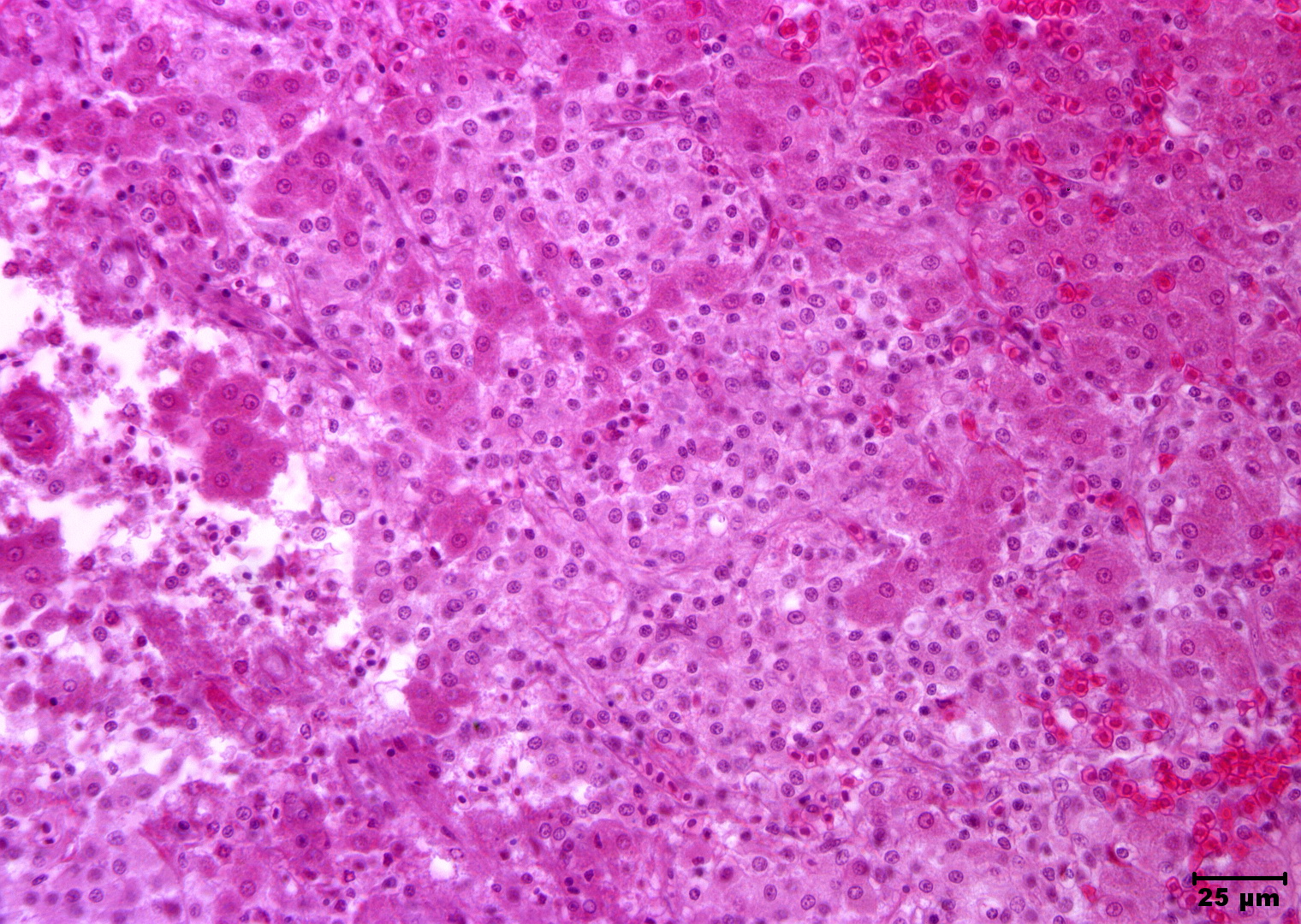
 | Slide # 33 Liver Hepatocellular Necrosis Edwardsiella ictaluri H&E 400X.jpg | All Pathogens, Fact Sheet Images | |
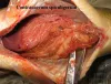
 | CB_BKD_01.jpg | All Pathogens, Fact Sheet Images | |
 | CB_BKD_05.jpg | All Pathogens, Fact Sheet Images | |
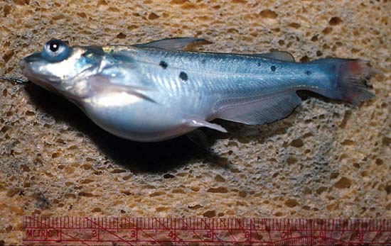
 | CCV-751.jpg | Fact Sheet Images | channel catfish |
 | IPN_Follasmolt_oerpetveit_web.jpg | Fact Sheet Images | Atlantic salmon |
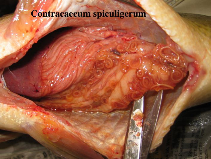
 | CB_Contracecum_01.jpg | All Pathogens, Fact Sheet Images | |
 | CB_BKD_04.jpg | All Pathogens, Fact Sheet Images | |
 | CB_BKD_03.jpg | All Pathogens, Fact Sheet Images | |
 | Neascus_05.jpg | All Pathogens, Fact Sheet Images | |