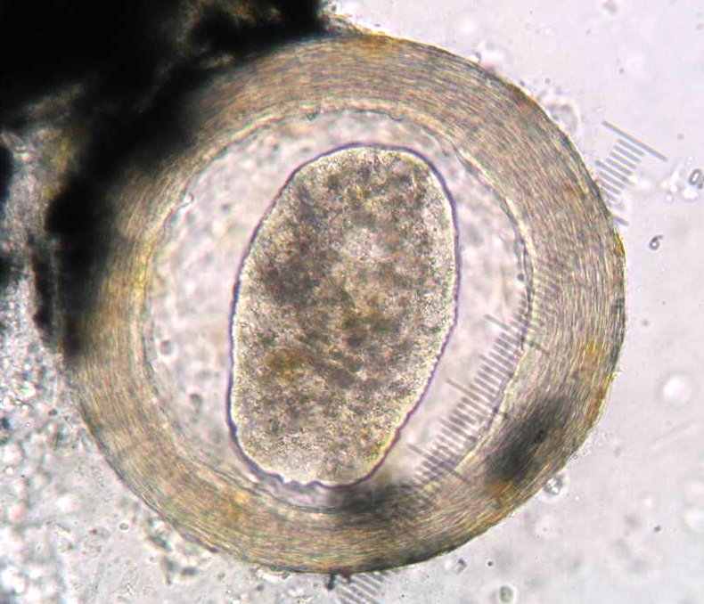
| Neascus_05.jpg | All Pathogens, Fact Sheet Images | |
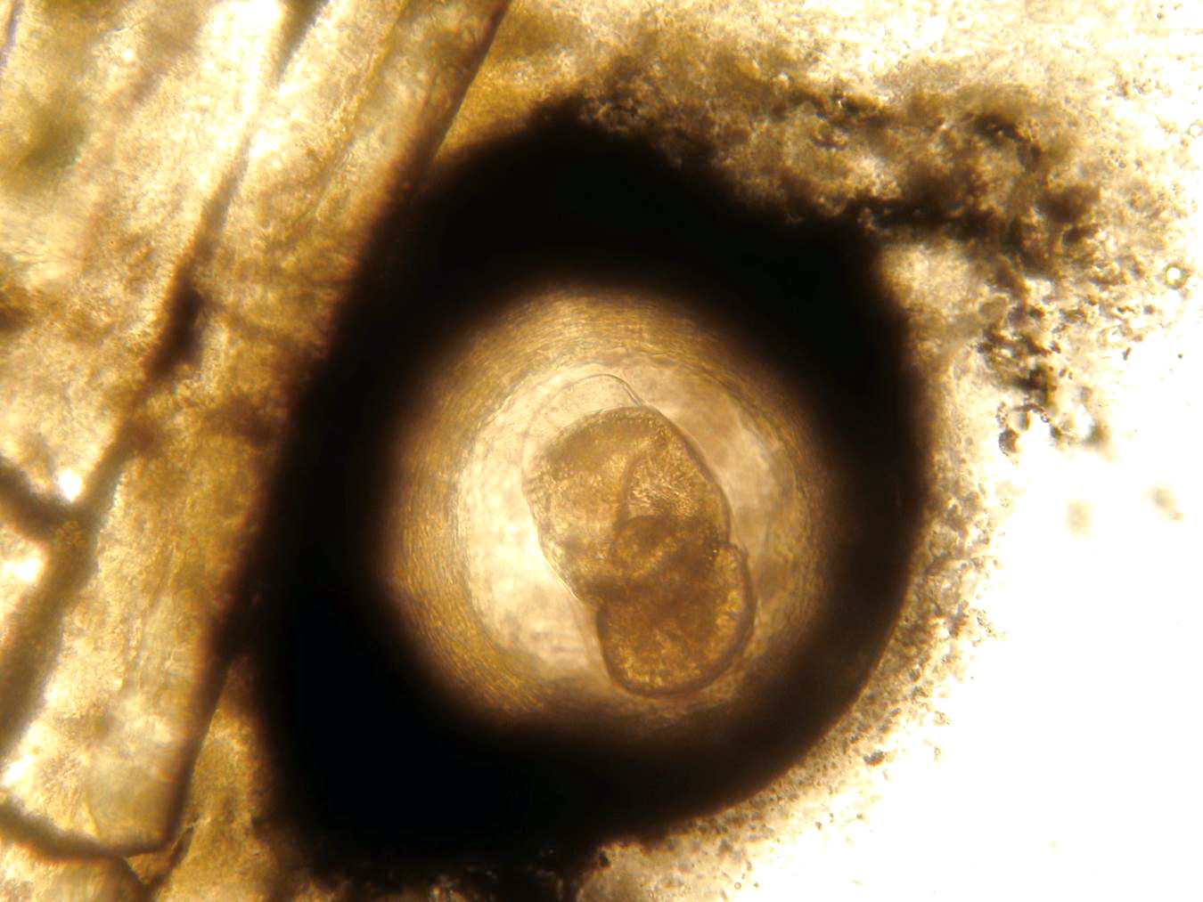
 | Neascus_06.jpg | All Pathogens, Fact Sheet Images | |
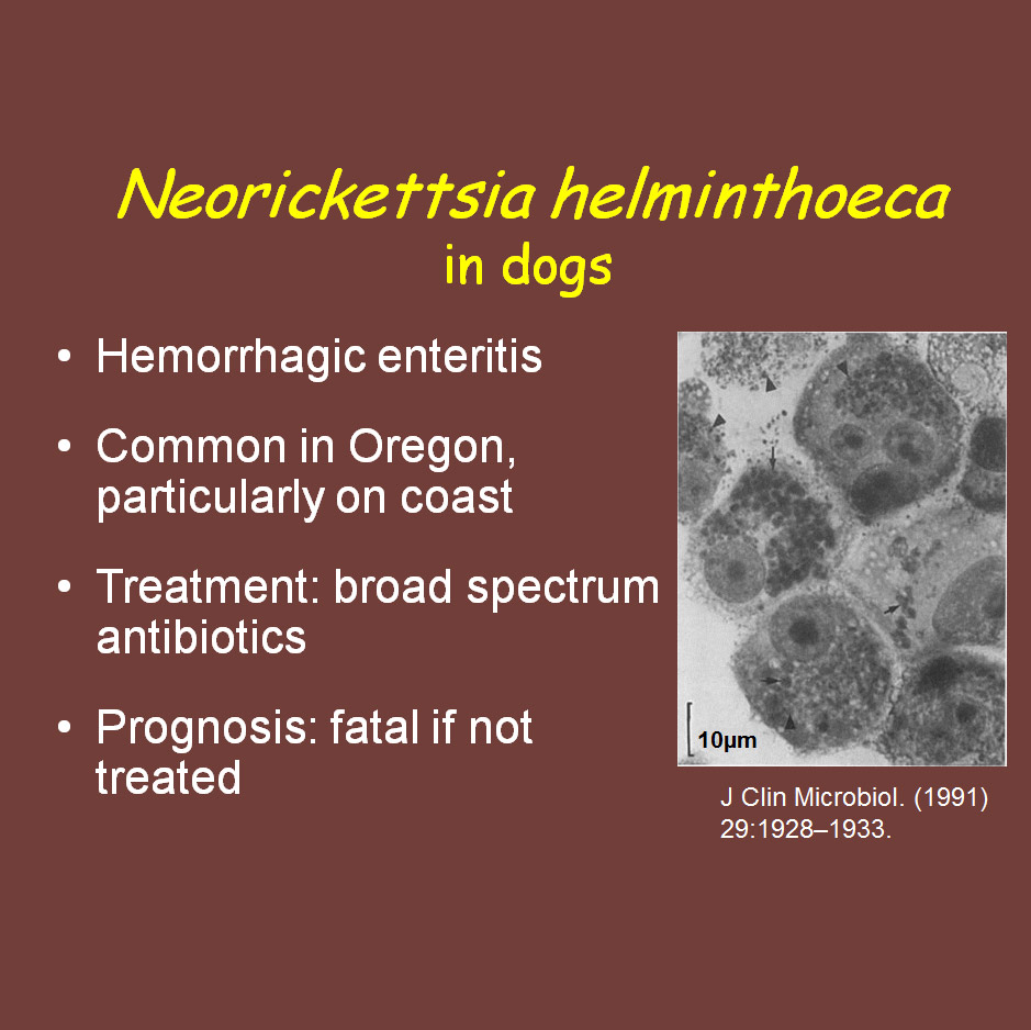
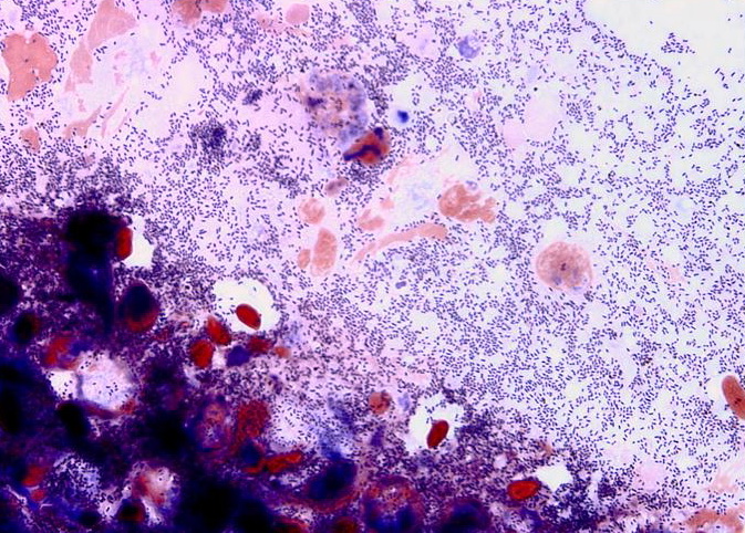
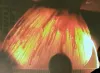
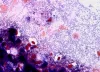
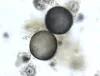
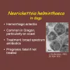 | CB_Neorickettsia_ppt.jpg | Fact Sheet Images, Illustration - organism | Nanophyetus |
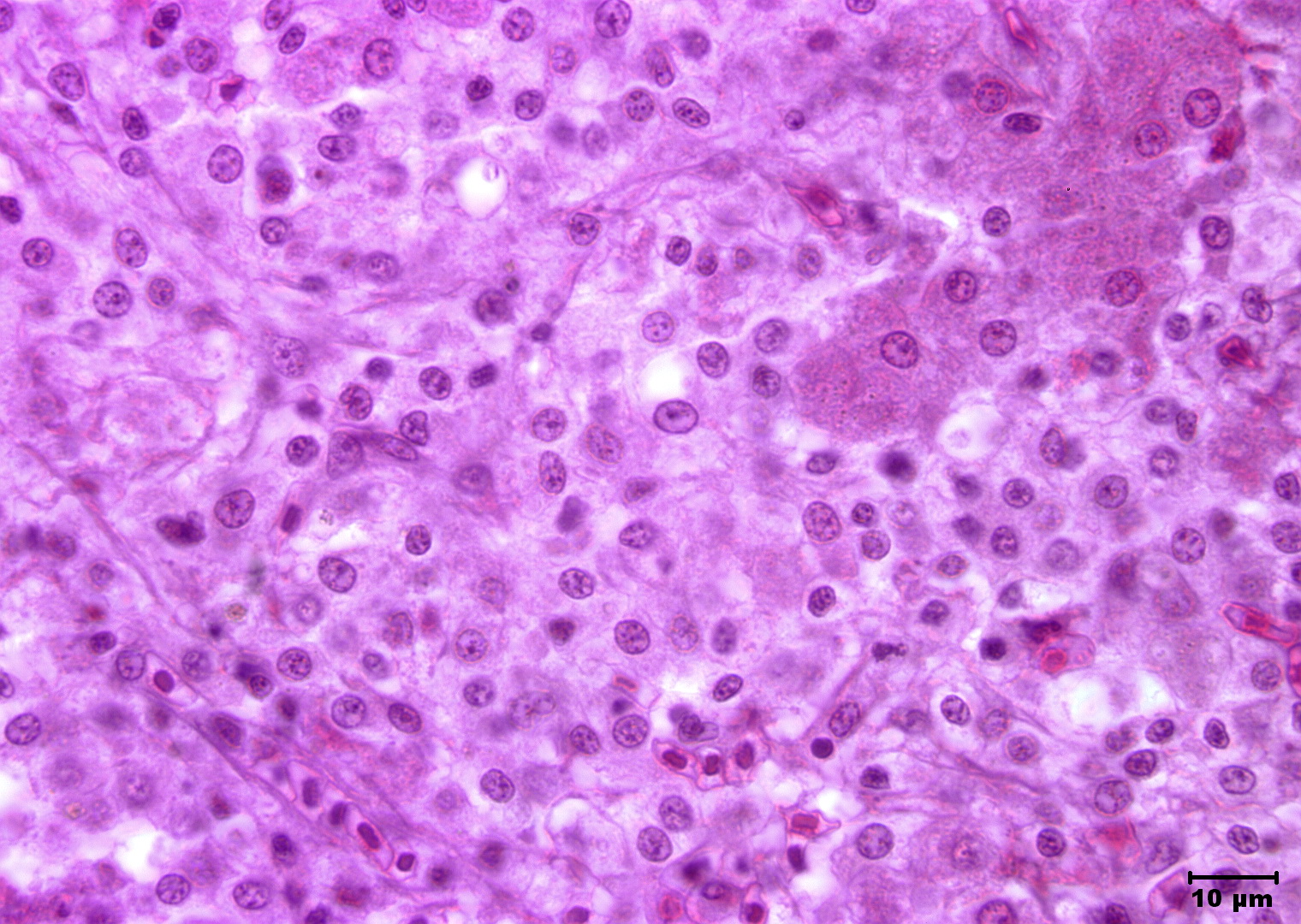
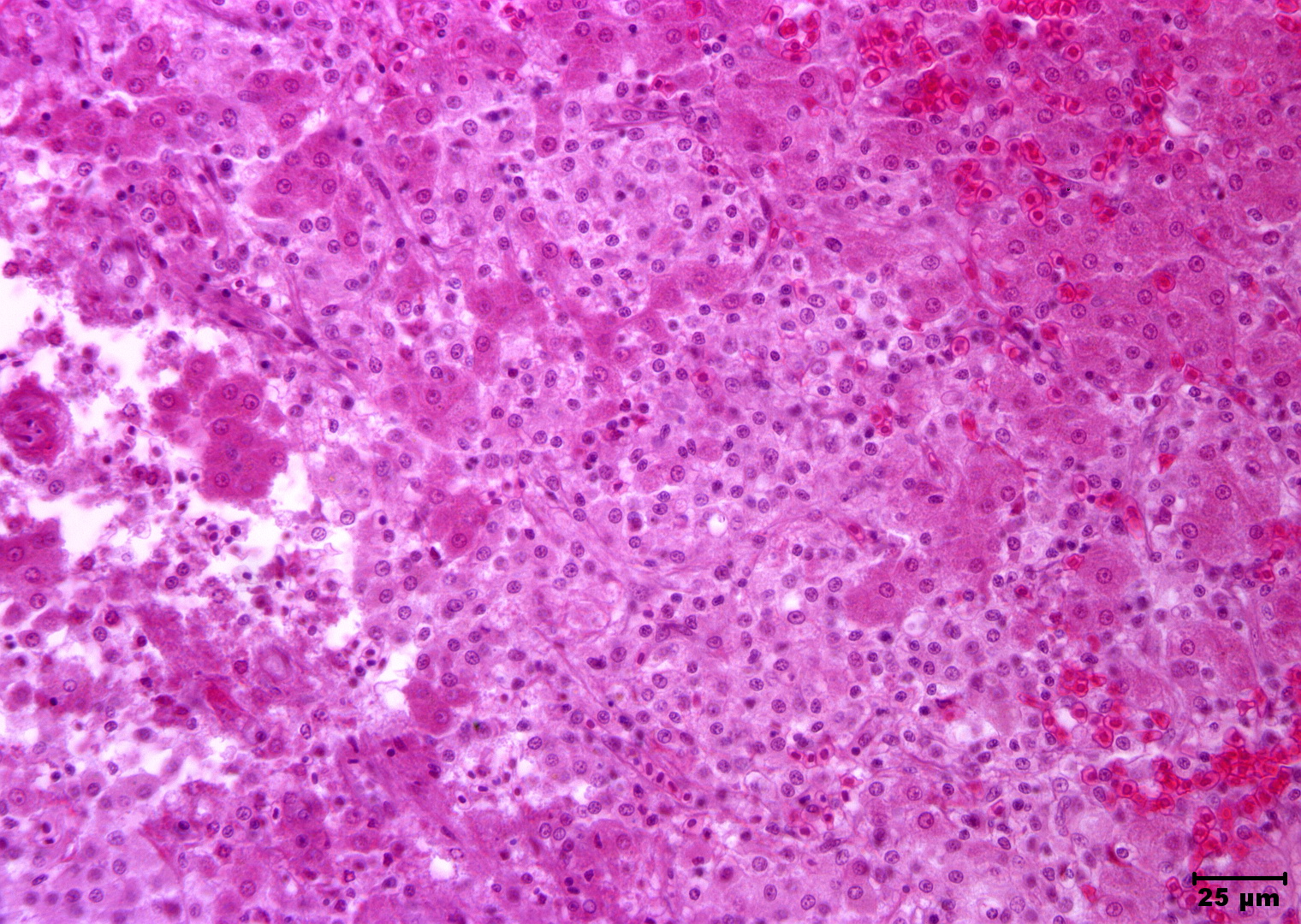
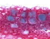
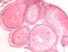
 | Slide # 33 Liver Hepatocellular Necrosis Edwardsiella ictaluri H&E Oil 1000X.jpg | All Pathogens, Fact Sheet Images | |
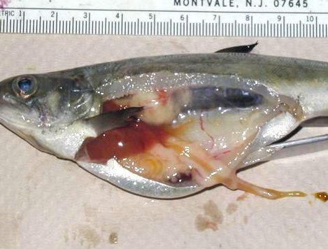
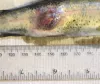
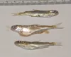
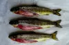
 | CB_Philometroides_01.jpg | All Pathogens, Fact Sheet Images | largescale sucker |
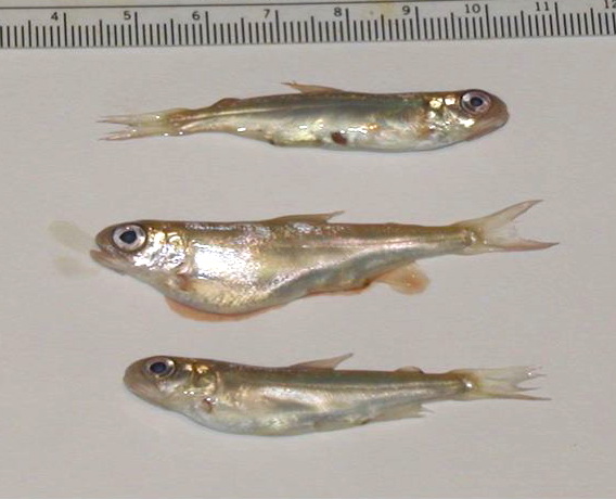
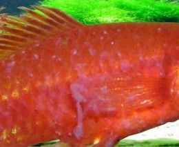
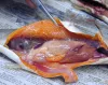
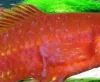
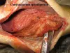
 | CB_Aeromonas_01.jpg | All Pathogens, Fact Sheet Images | rainbow trout |
 | Slide # 33 Liver Hepatocellular Necrosis Edwardsiella ictaluri H&E 400X.jpg | All Pathogens, Fact Sheet Images | |
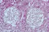
 | CB_BKD_01.jpg | All Pathogens, Fact Sheet Images | |
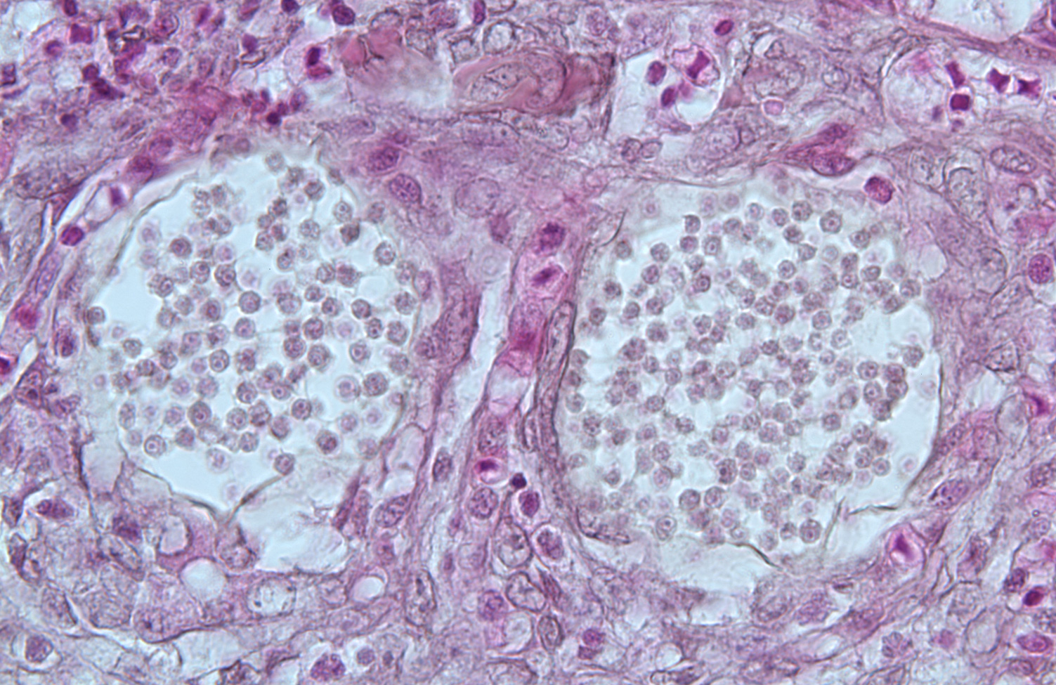
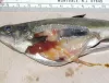 | CB_BKD_02_ChS.jpg | All Pathogens, Fact Sheet Images | Spring Chinook salmon |
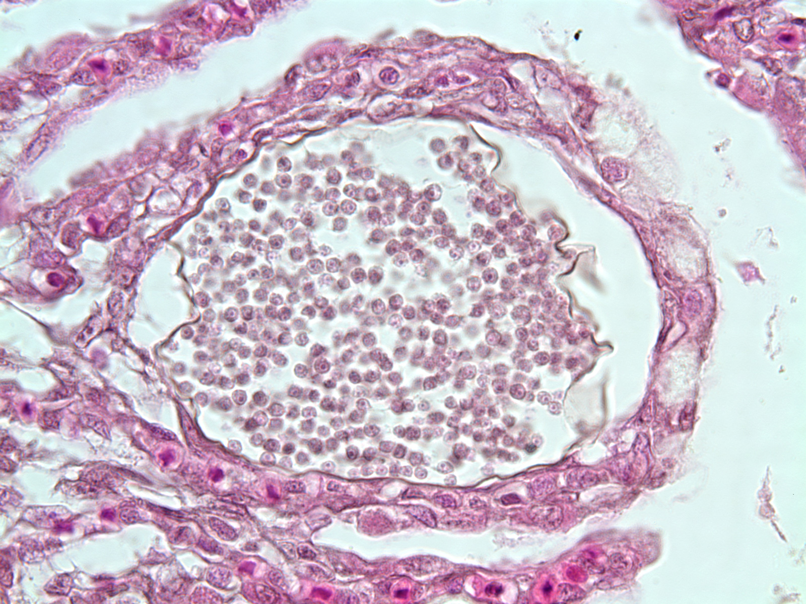
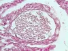
 | CB_BKD_03.jpg | All Pathogens, Fact Sheet Images | |
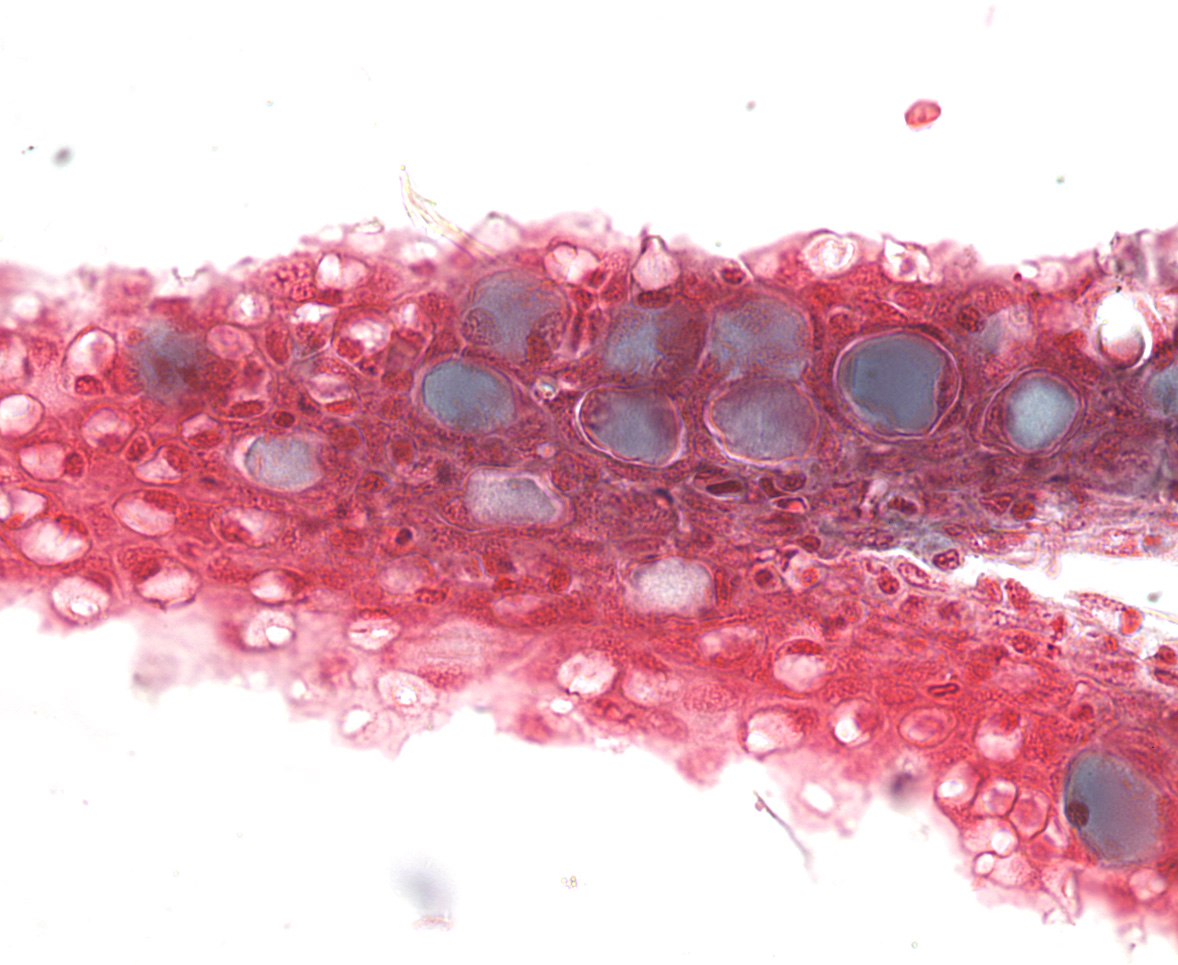
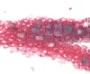
 | CB_BKD_04.jpg | All Pathogens, Fact Sheet Images | |
 | 1603391_f260_velvet.jpg | Fact Sheet Images | |
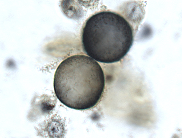
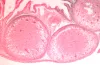
 | CB_BKD_05.jpg | All Pathogens, Fact Sheet Images | |
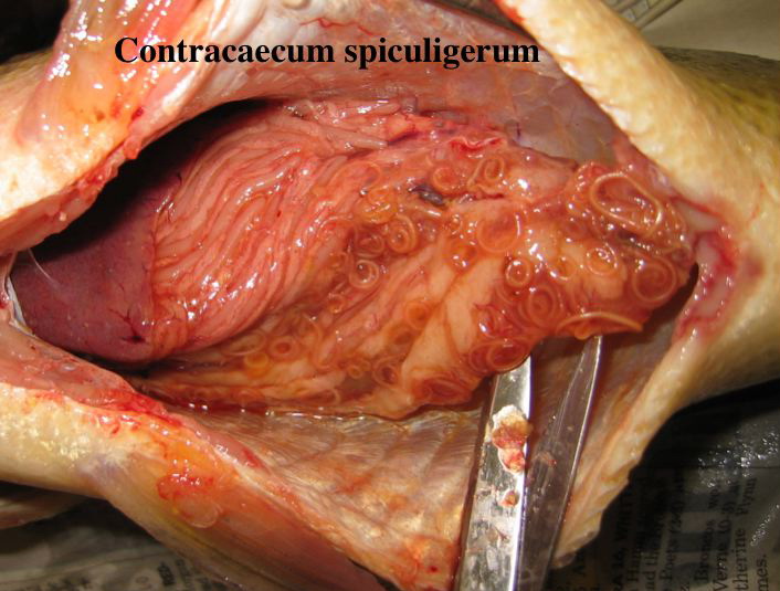
 | CB_Contracecum_01.jpg | All Pathogens, Fact Sheet Images | |
 | IPN_Follasmolt_oerpetveit_web.jpg | Fact Sheet Images | Atlantic salmon |
 | CCV-751.jpg | Fact Sheet Images | channel catfish |
 | BO_Ich_x63_10.jpg | All Pathogens, Fact Sheet Images | |
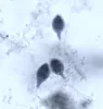
 | BO_Ichthyobodo_x63_32.jpg | All Pathogens, Fact Sheet Images | Chinook salmon |
 | BO_Dermocystidium_x63_24.jpg | All Pathogens, Fact Sheet Images | |
 | BO_Dermocystidium_x63_25.jpg | All Pathogens, Fact Sheet Images | |
 | BO_Ichthyobodo_x63_31.jpg | All Pathogens, Fact Sheet Images | Chinook salmon |
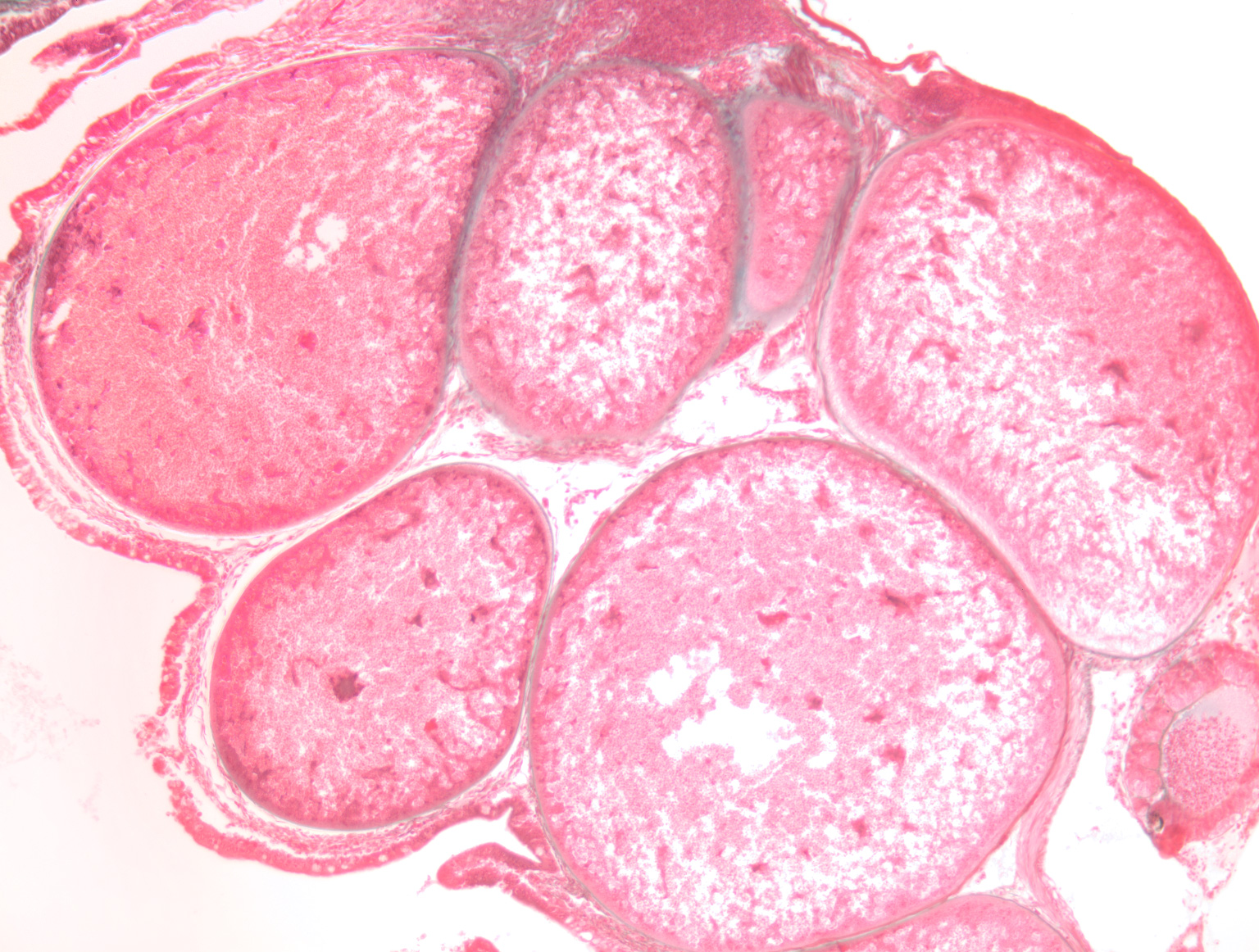
 | BO_Glugea_x10_14.jpg | All Pathogens, Fact Sheet Images | |
 | BO_Ichthyobodo_x63_29.jpg | All Pathogens, Fact Sheet Images | Chinook salmon |
 | BO_Glugea_x10_15.jpg | All Pathogens, Fact Sheet Images | |
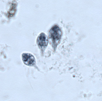
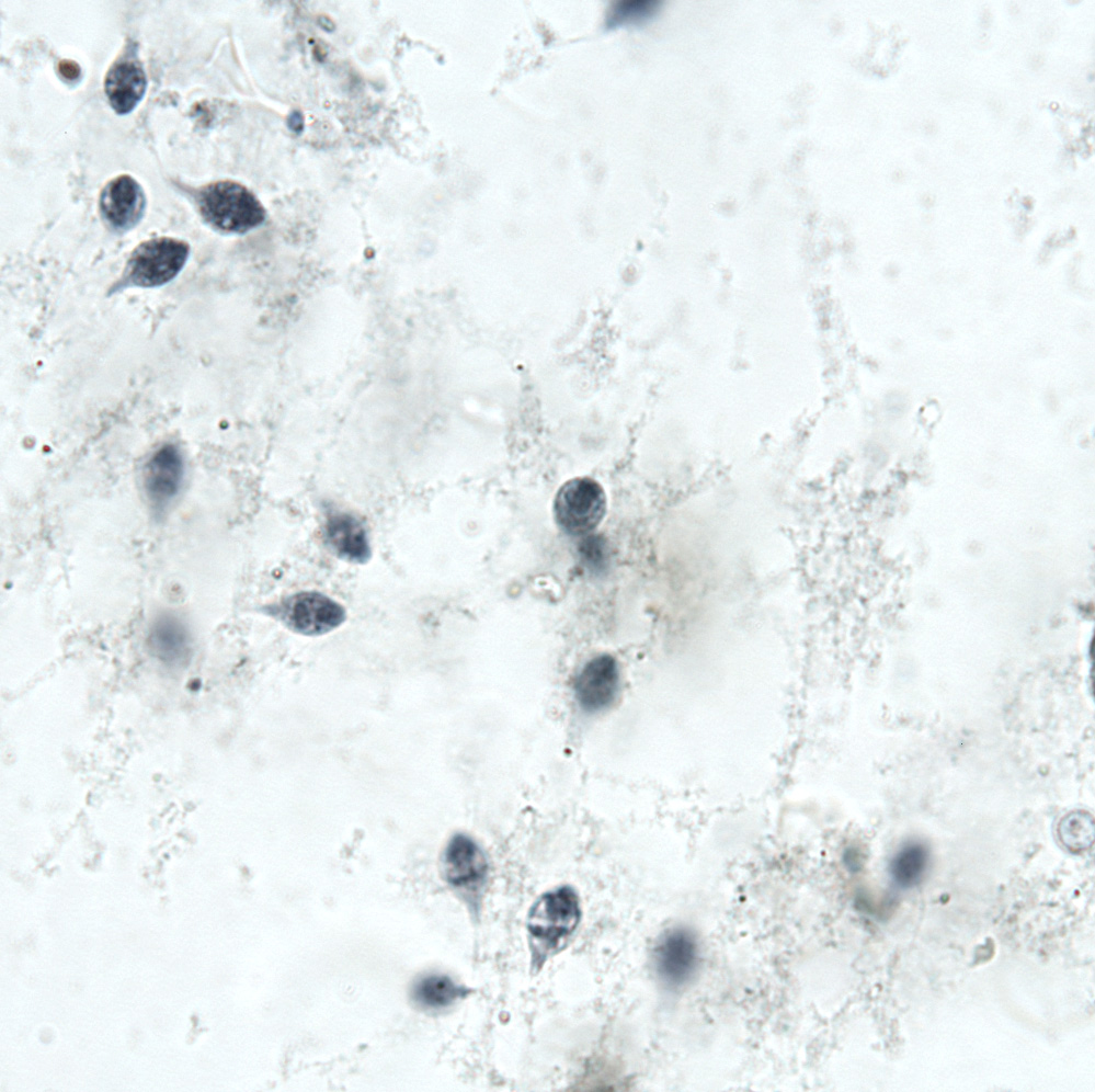
 | BO_Hexamita_x63_36.jpg | All Pathogens, Fact Sheet Images | |
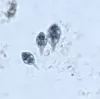 | BO_Hexamita_x63_35.jpg | All Pathogens, Fact Sheet Images | |
 | BO_Hexamita_x63_34.jpg | All Pathogens, Fact Sheet Images | |
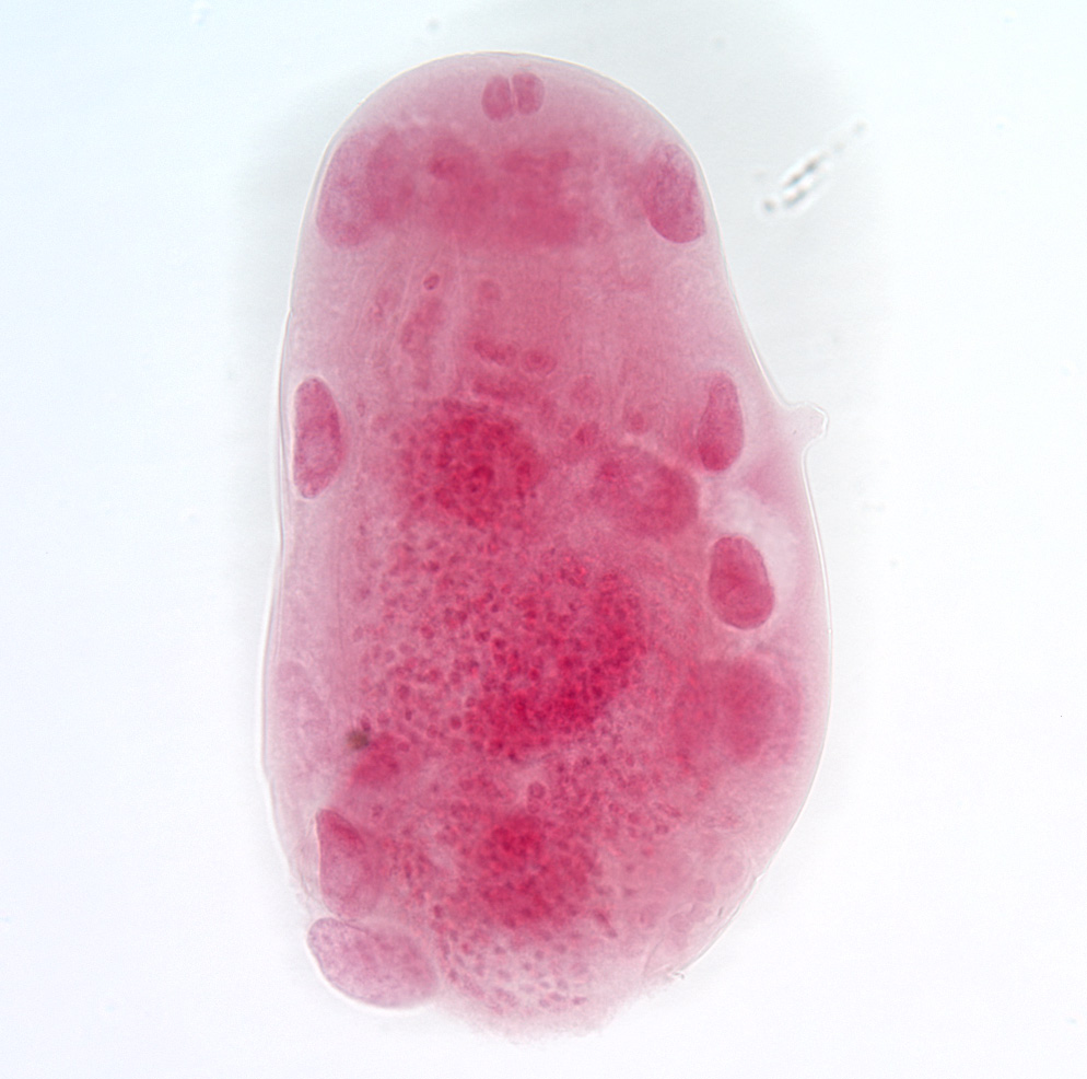
 | BO_Echinorhynchus_x20_27.jpg | All Pathogens, Fact Sheet Images | amphipod |
 | BO_Ich_x63_09.jpg | All Pathogens, Fact Sheet Images | |
 | BO_Glugea_x63_16.jpg | All Pathogens, Fact Sheet Images | |