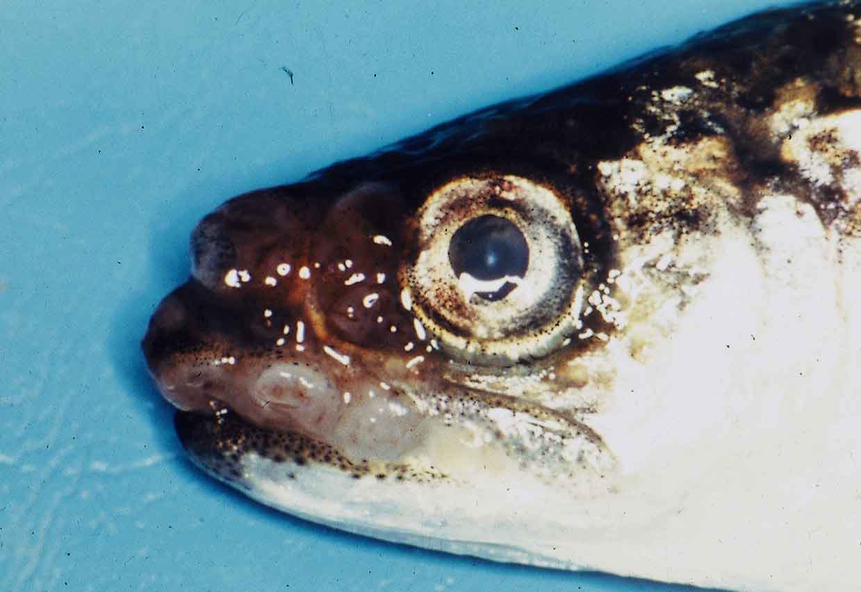
 | OMV-Masu.jpg | All Pathogens, Fact Sheet Images | |
 | sanguinicola_CB08.jpg | All Pathogens, Fact Sheet Images | |
 | Cshasta03.jpg | All Pathogens, Fact Sheet Images | |
 | sanguinicola_CB07.jpg | All Pathogens, Fact Sheet Images | |
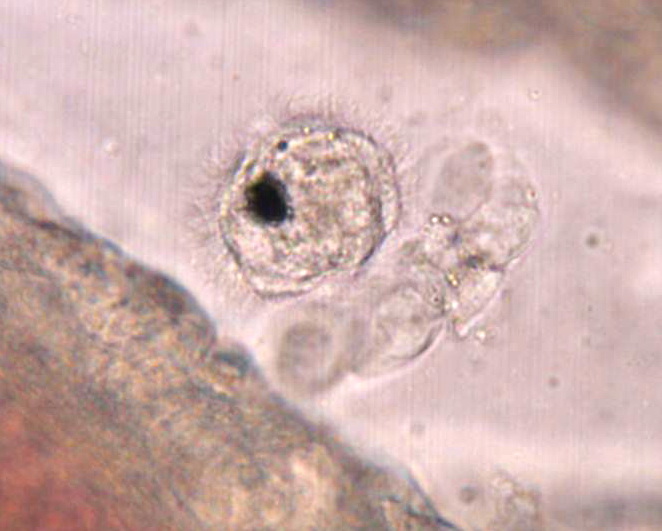
 | BO_miracidium_x63_13.jpg | All Pathogens | |
 | sanguinicola_CB06.jpg | All Pathogens, Fact Sheet Images | |
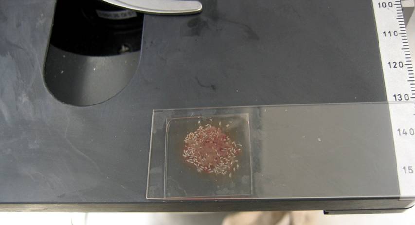
 | 177_7781.JPG | All Pathogens, Fact Sheet Images | |
 | BO_miracidium_x63_12.jpg | All Pathogens | |
 | IMG_20130402_rbtCs2.jpg | All Pathogens | rainbow trout |
 | CB_IMG_0352.jpg | All Pathogens, Fact Sheet Images, Top 10, Hosts and other organisms | brown trout |
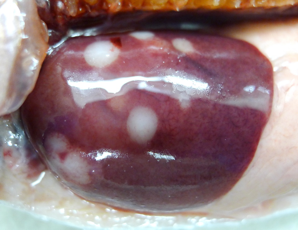
 | P1080678_Cshasta_liver.jpg | All Pathogens | rainbow trout |
 | sanguinicola_CB04.jpg | All Pathogens, Fact Sheet Images | |
 | CB_Kidtremyellow.jpg | All Pathogens, Fact Sheet Images | bluegill |
 | BO_Loma_x63_20.jpg | All Pathogens | |
 | sanguinicola_CB01_miracidium.jpg | All Pathogens, Fact Sheet Images | |
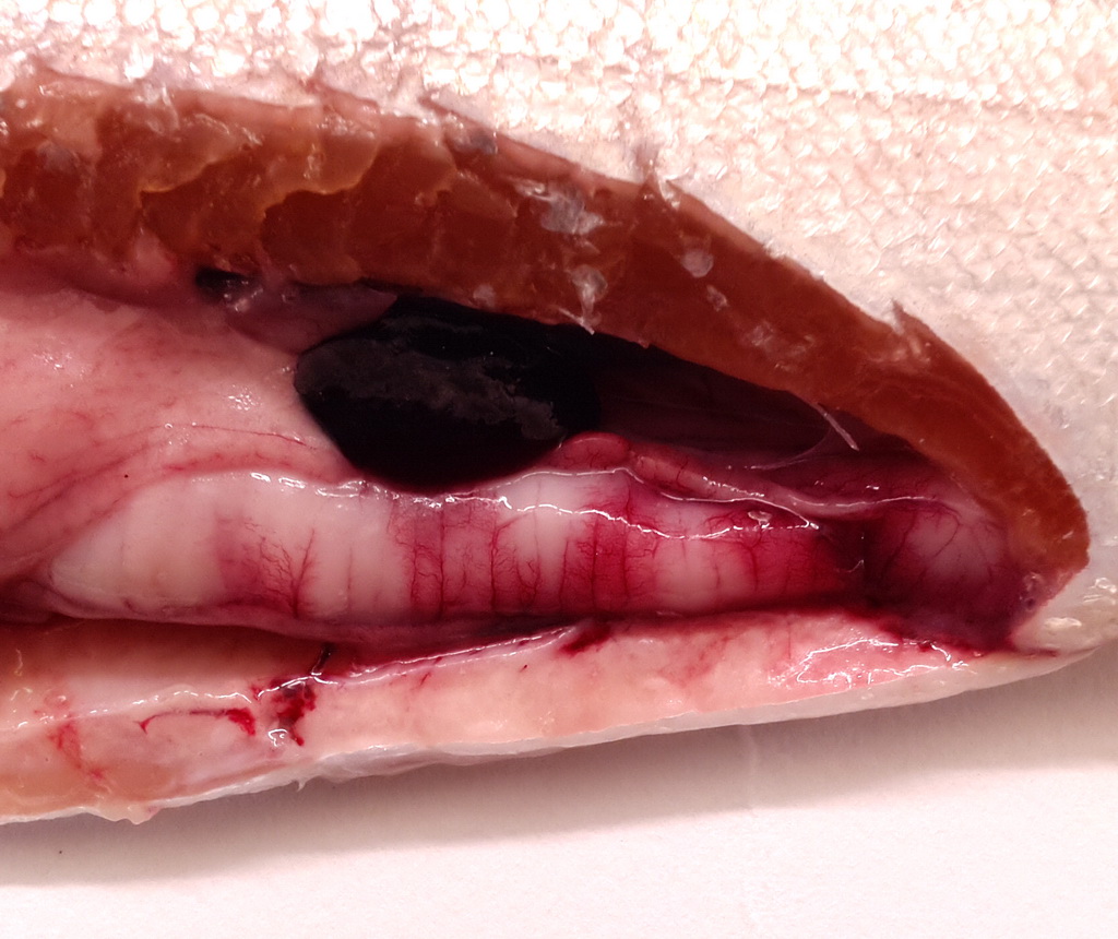
 | P1070955_Cshasta.jpg | All Pathogens | rainbow trout |
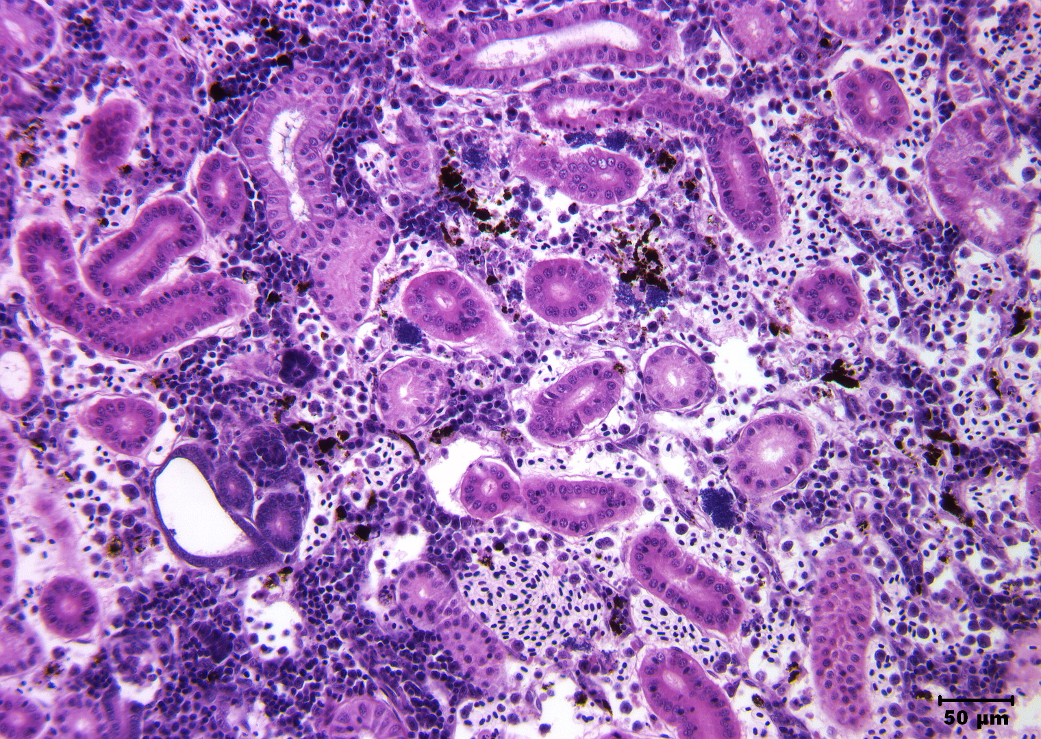
 | Slide # 18 Kidney Aeromonas salmonicida H&E 200X.jpg | All Pathogens | |
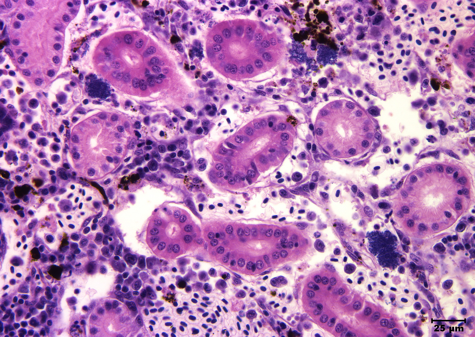
 | Slide # 18 Kidney Aeromonas salmonicida H&E 400X.jpg | All Pathogens | |
 | salmincola-CB04.jpg | All Pathogens, Fact Sheet Images | |
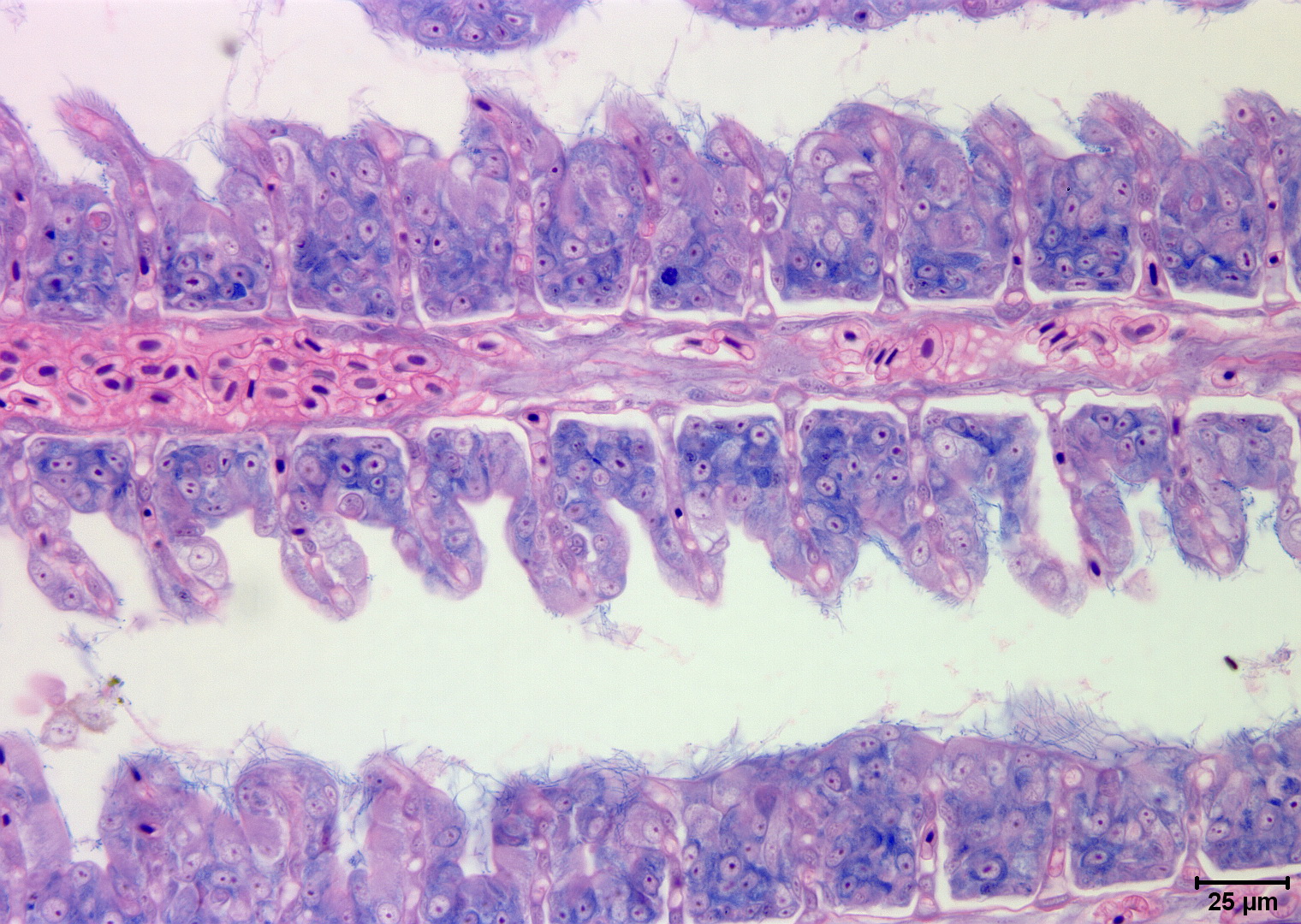
 | Slide # 37 Gills Flavobacterium branchiophilus Giemsa 400X.jpg | All Pathogens | |
 | Clinostomum_05.jpg | All Pathogens, Fact Sheet Images | |
 | Slide # 40 Necrotizing steatitis Flavobacterium psychrophilus H&E 400X.jpg | All Pathogens, Fact Sheet Images | |
 | nanophyetus_salmonicola_CB07.jpg | All Pathogens, Fact Sheet Images | |
 | Clinostomum_02.jpg | All Pathogens, Fact Sheet Images | |
 | nanophyetus_salmonicola_CB08.jpg | All Pathogens, Fact Sheet Images | |
 | BO_Ichthyophonus_x20_37.jpg | All Pathogens | |
 | nanophyetus_salmonicola_CB09.jpg | All Pathogens, Top 10, Fact Sheet Images | yellow perch |
 | BO_Ichthyophonus_x20_38.jpg | All Pathogens | |
 | Slide # 36 Gills Flavobacterium branchiophilus H&E 400X.jpg | All Pathogens | |
 | Slide # 36 Gills Flavobacterium branchiophilus H&E 400X 2.jpg | All Pathogens | |