Skip to main content
Fish Pathogens
Menu
Search
Search
Main navigation
Image Galleries
Pathogens
Pathogens sub-navigation
10 Common Pathogens
What's That Spot?
Ask an Expert
FAQ
Search
Search
Search
User account menu
Log in
Primary tabs
Common Pathogens
All Pathogens
Hosts
Field Sites
Videos
Contributed
All Pathogens
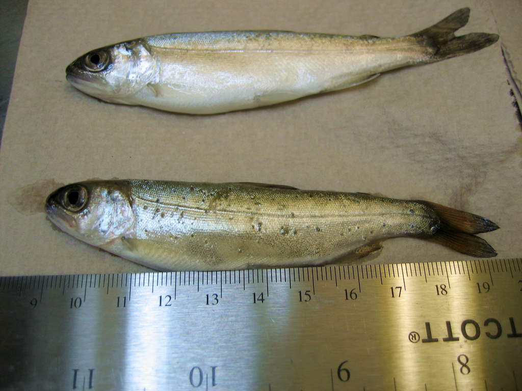
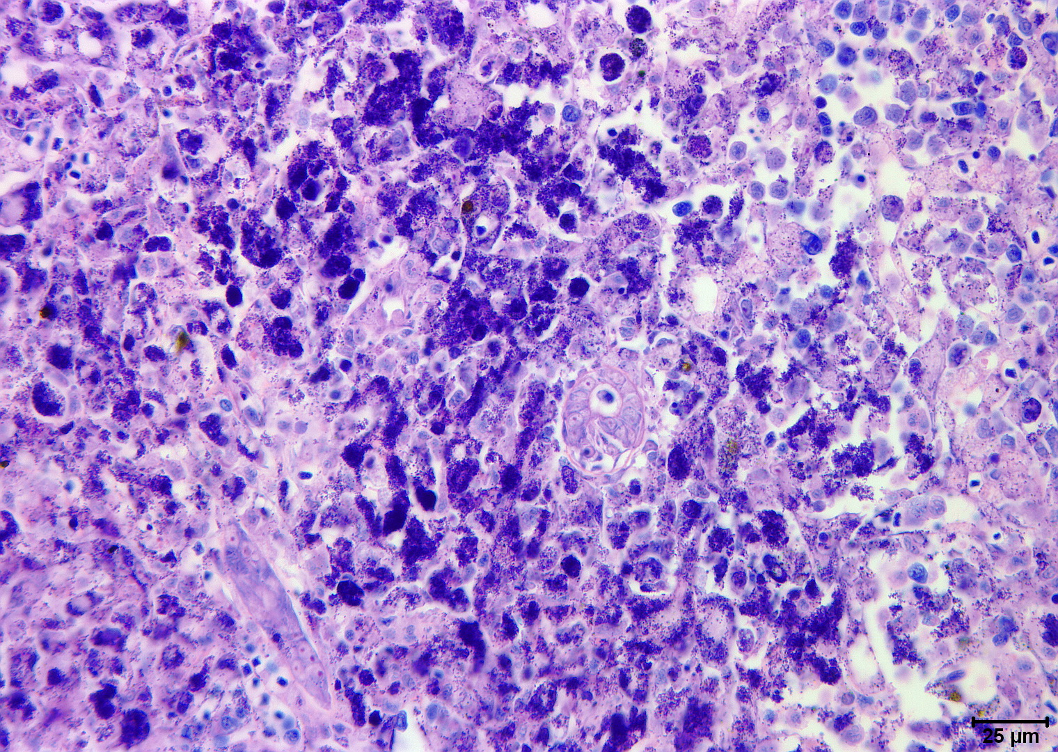
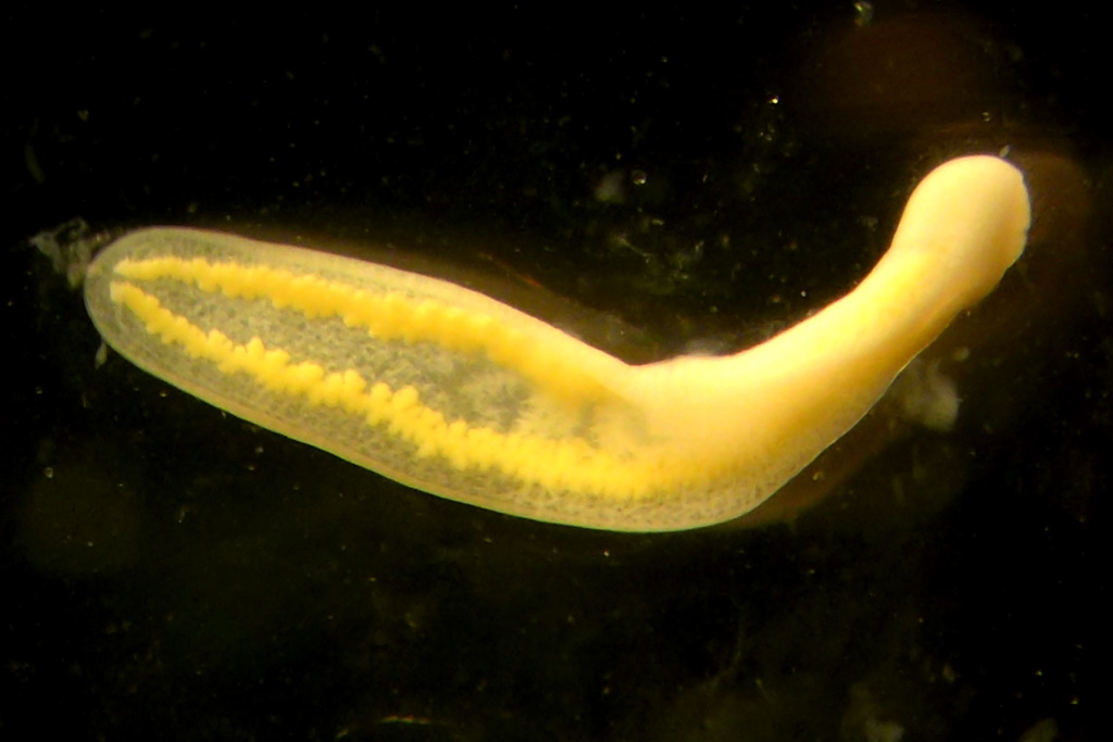
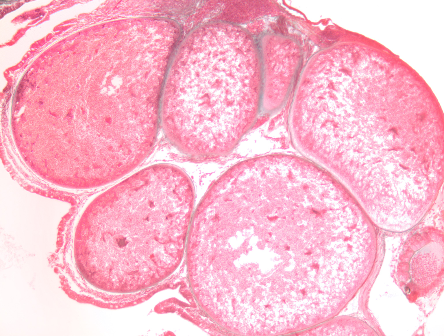
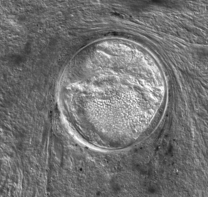
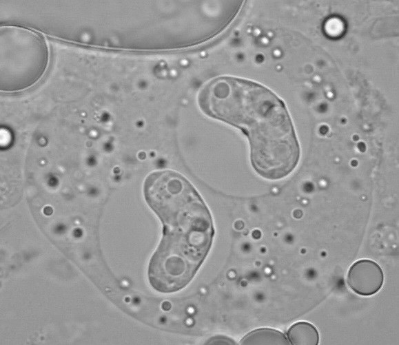
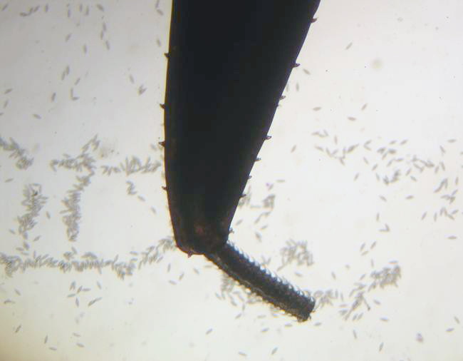
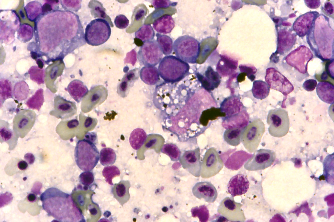
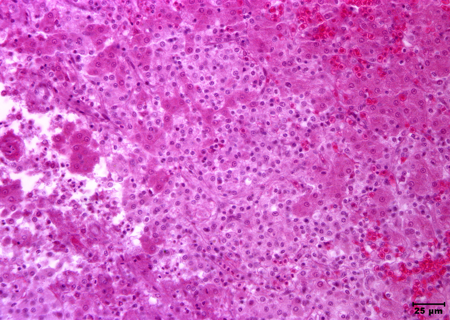
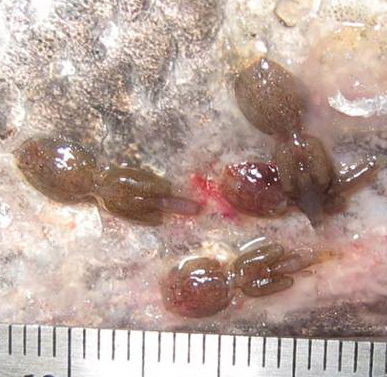
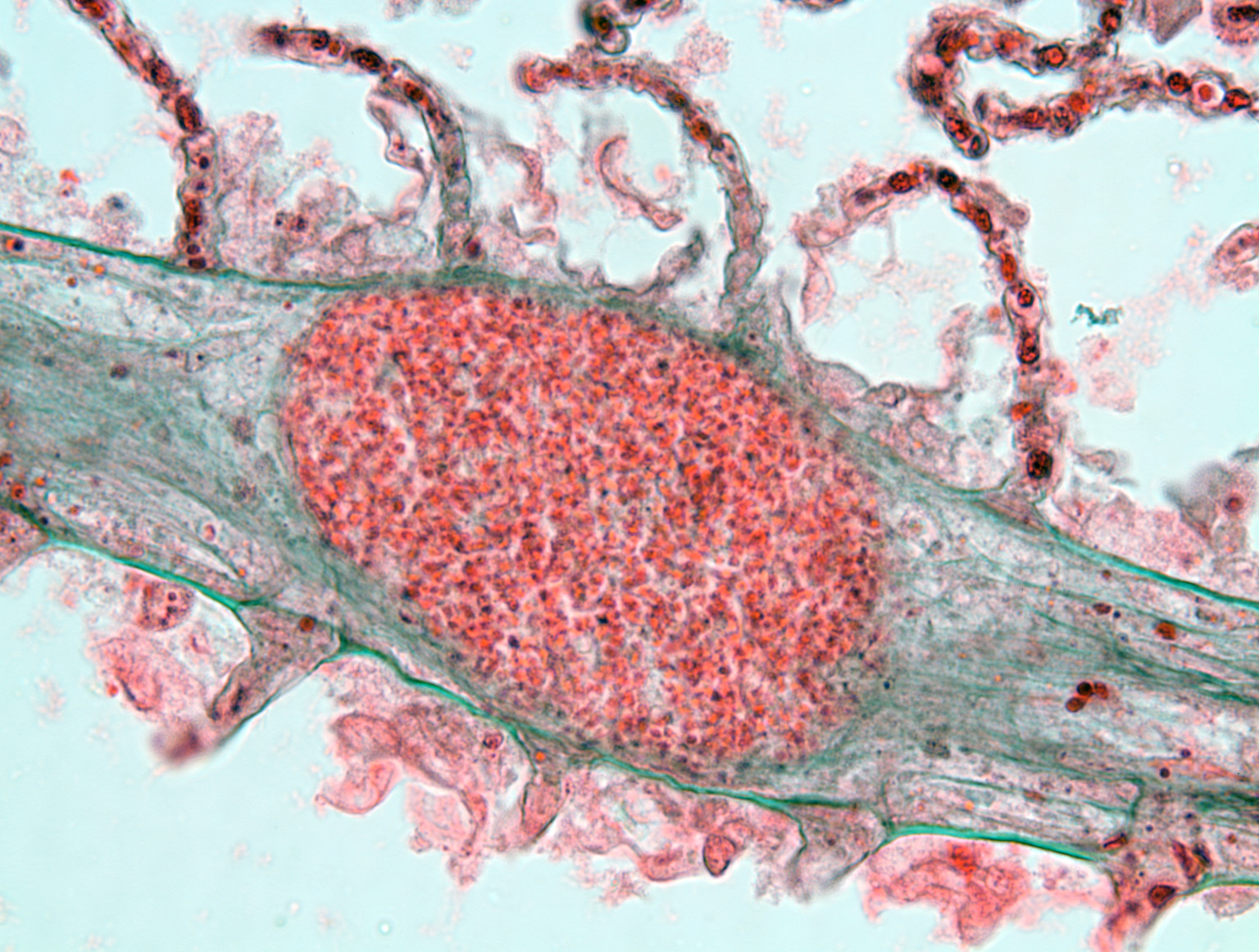
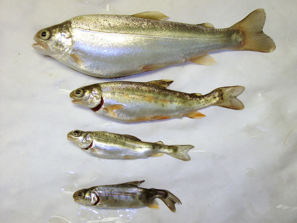
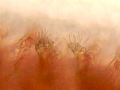
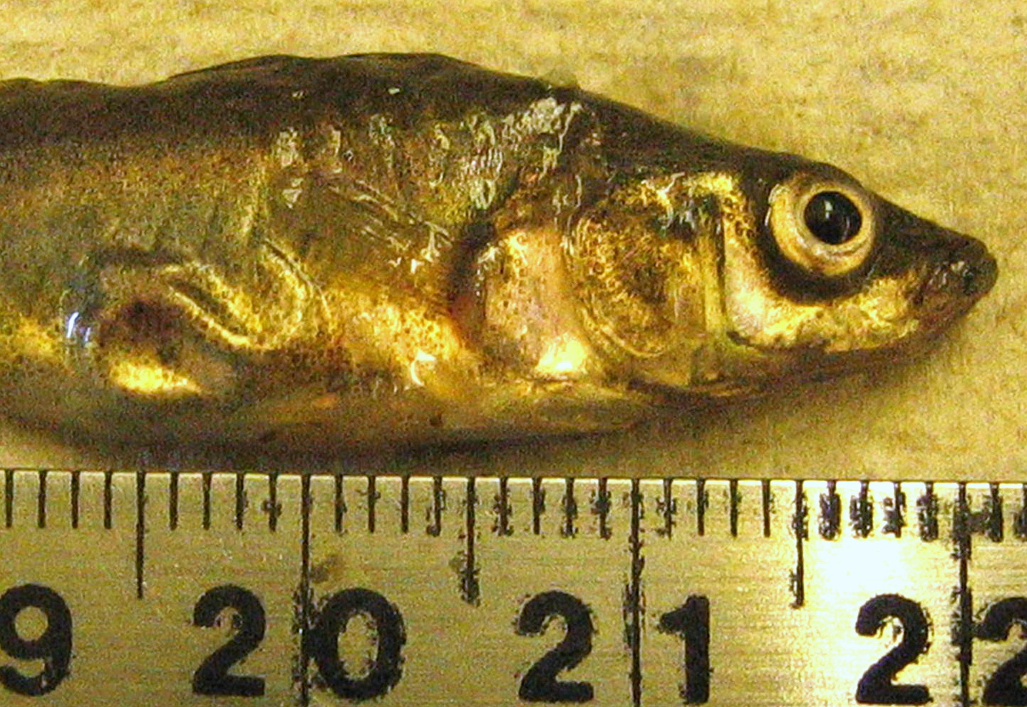
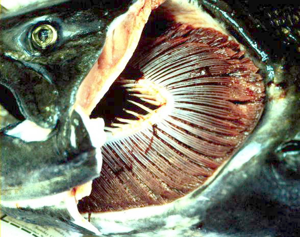
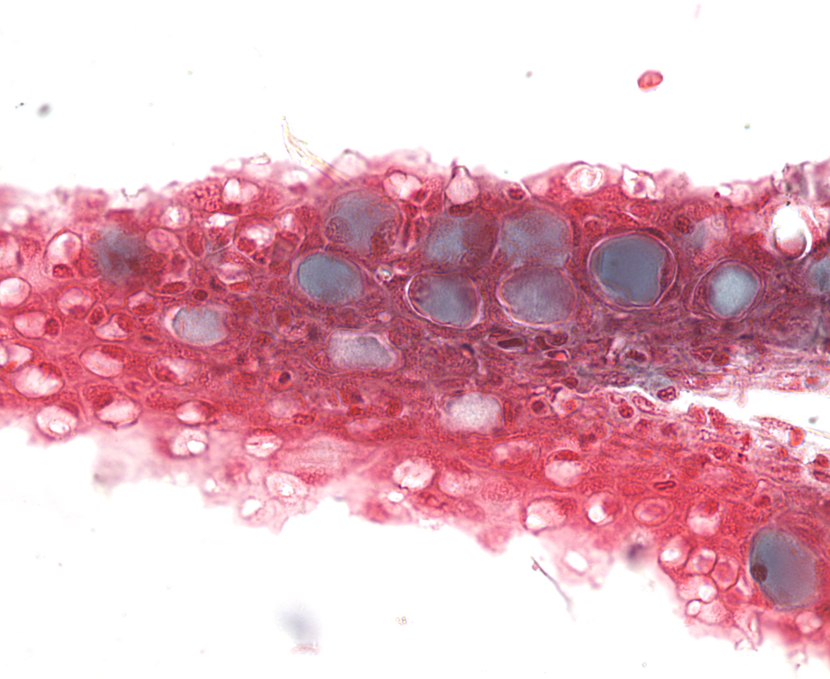
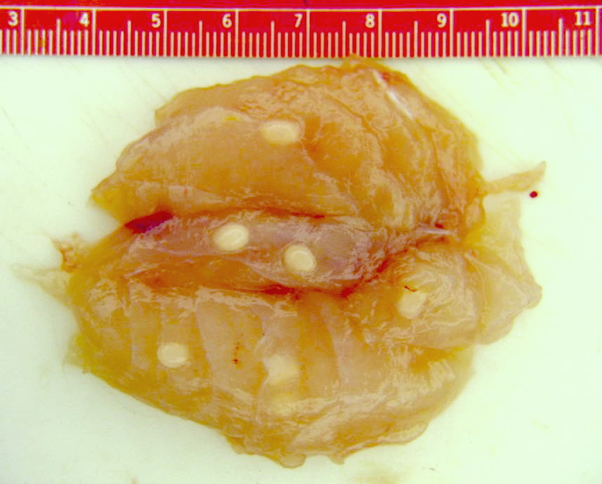
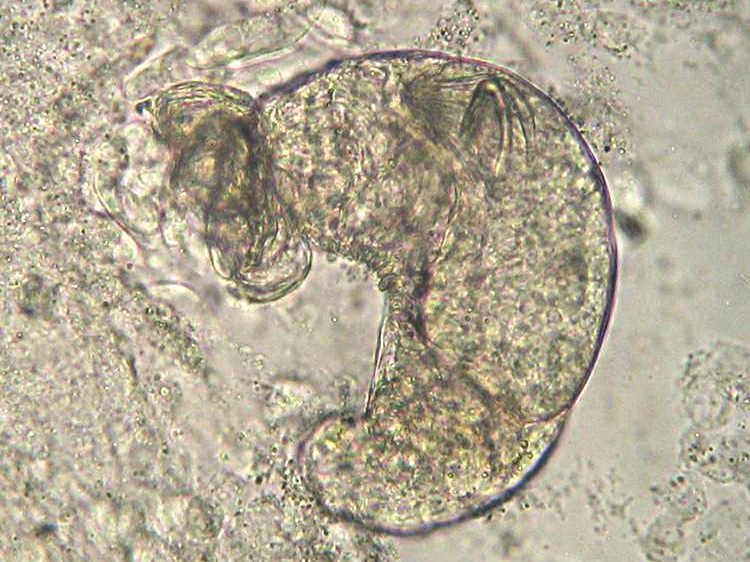
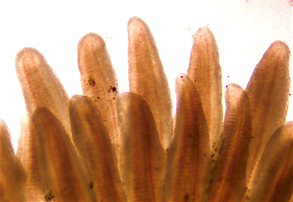
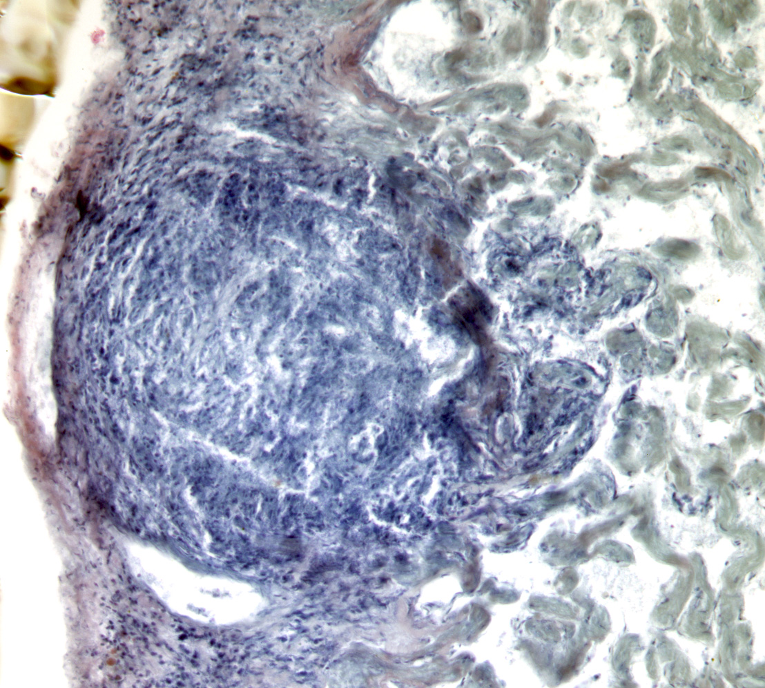
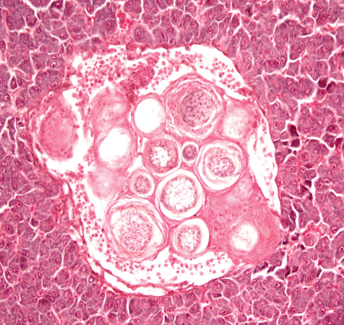
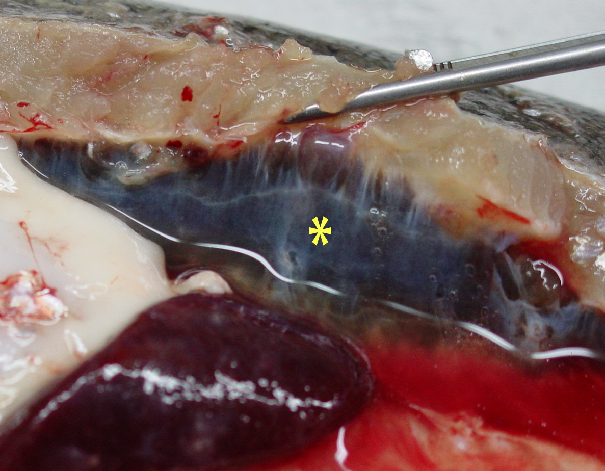
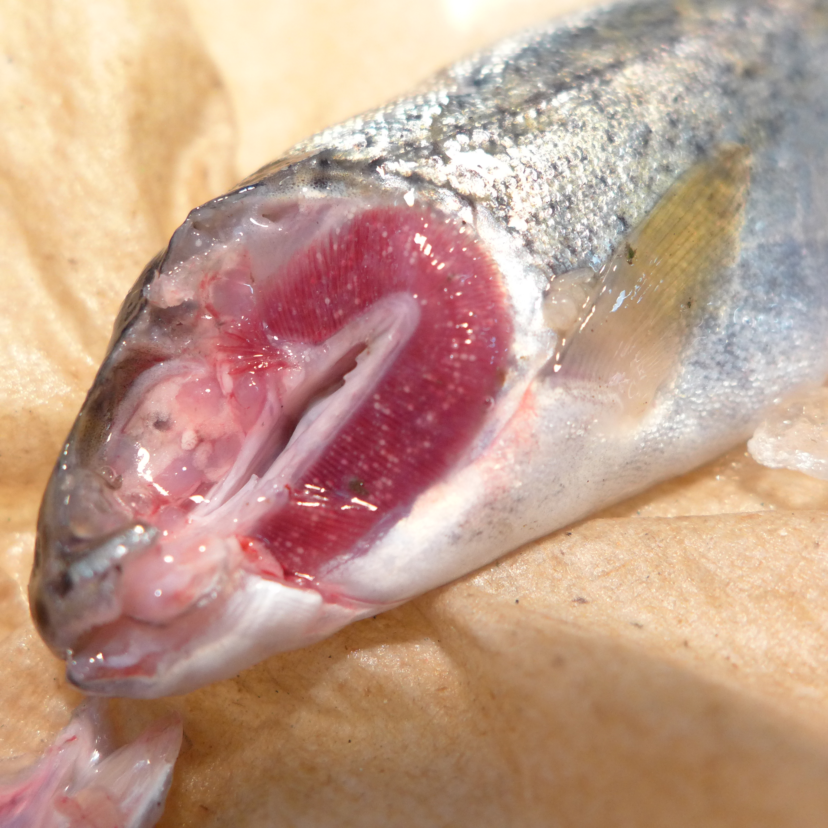
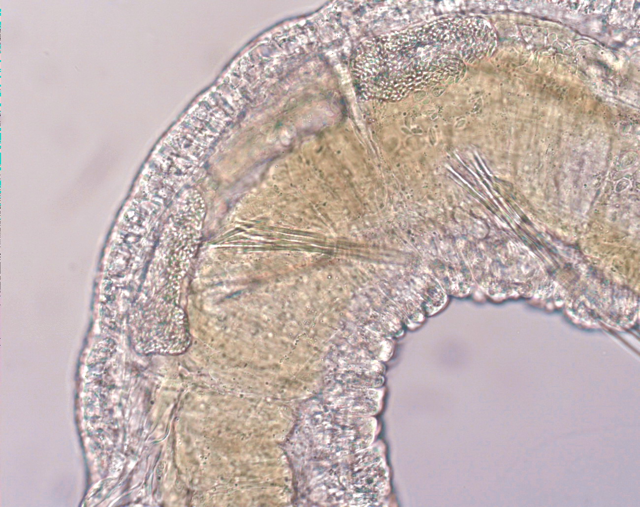
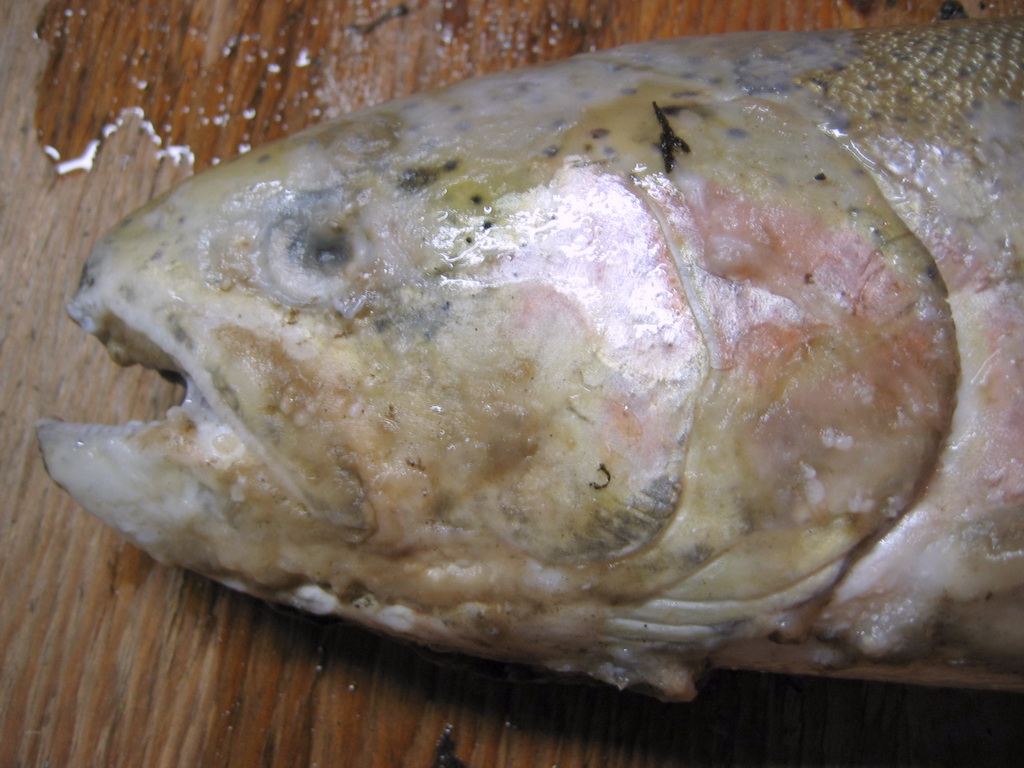
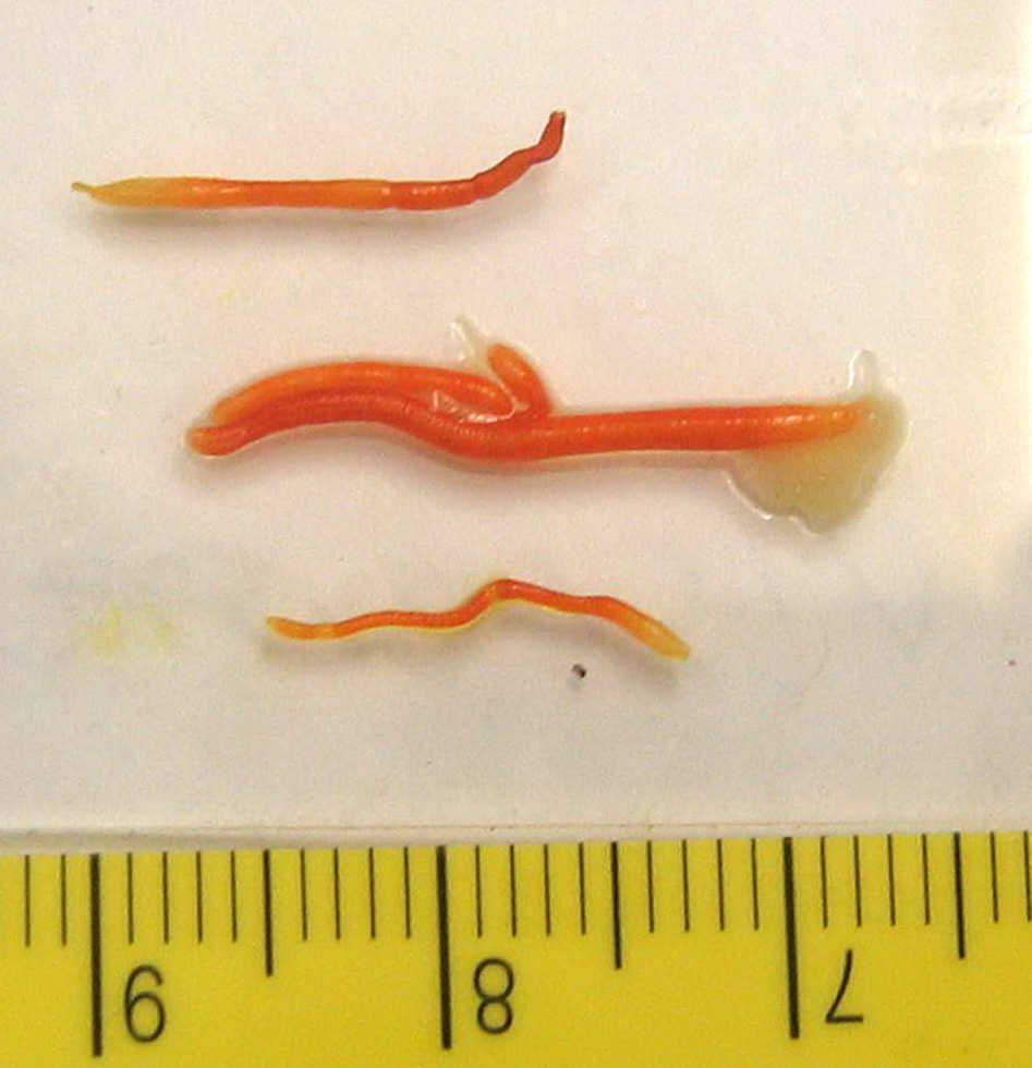
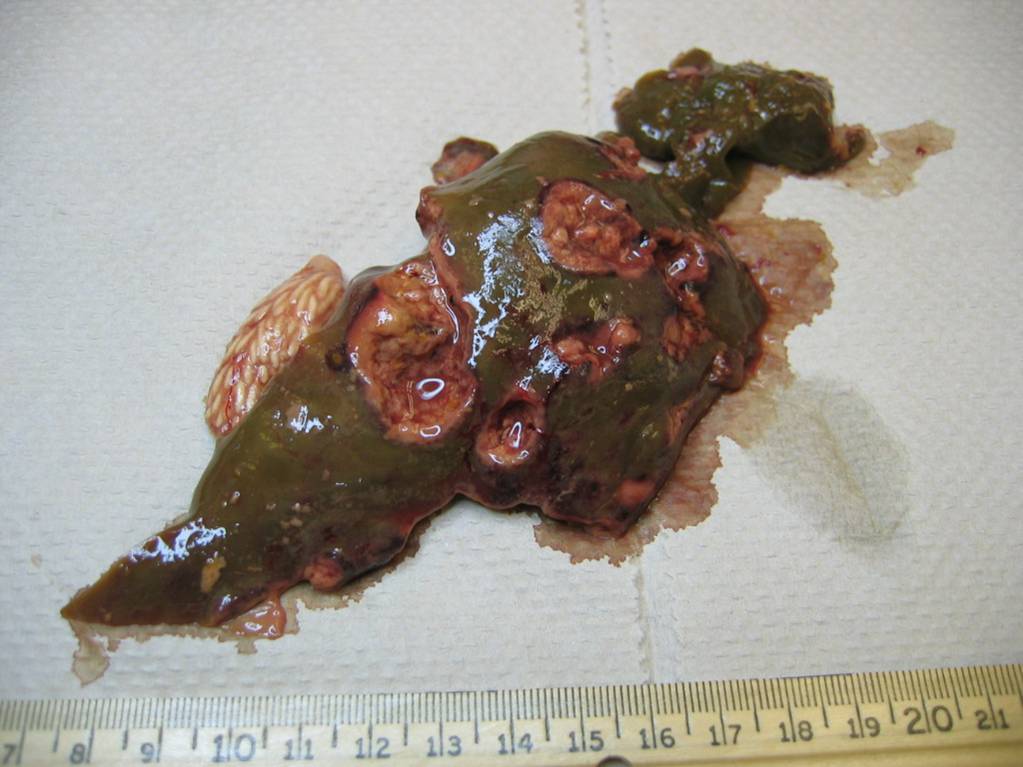
These are all of the pathogen images in our database. Click a pathogen name to jump to the pathogen info page. Click an image to enlarge it, then use the left and right arrows to browse.
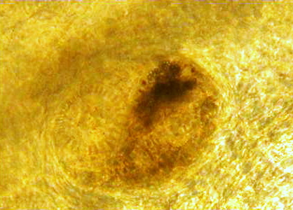
Clinostomum (Yel…
Myxobolus cerebrali…
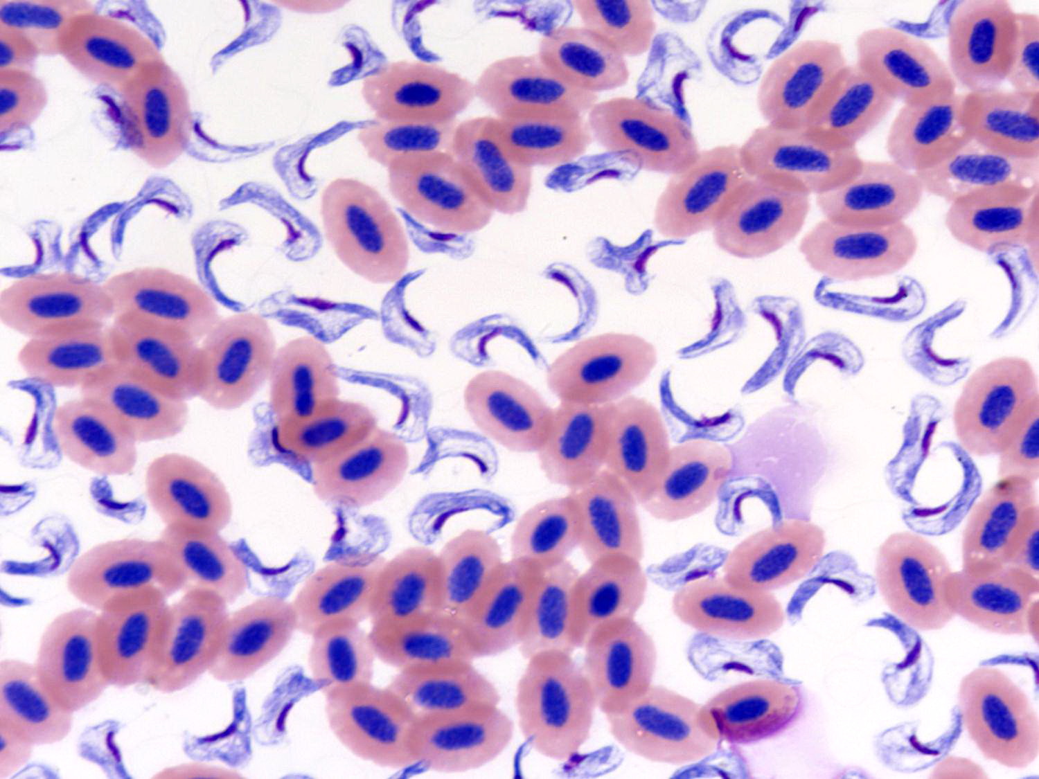
Cryptobia salmosit…
Myxobolus insidiosu…
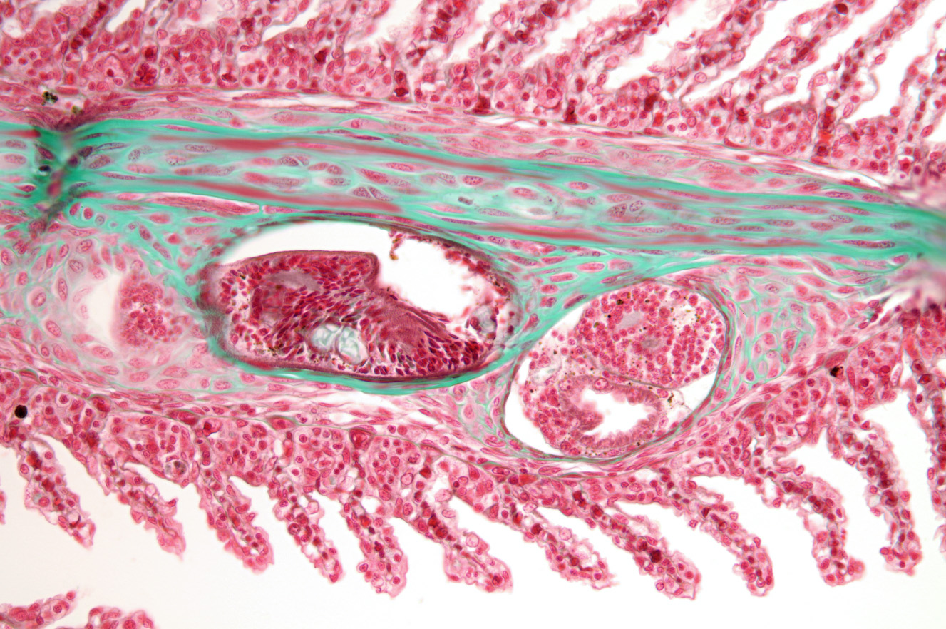
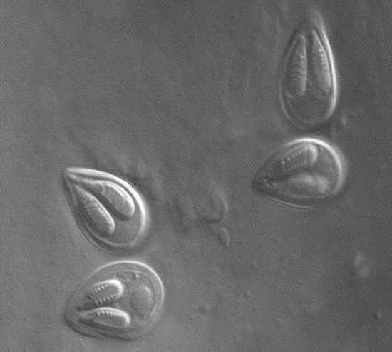
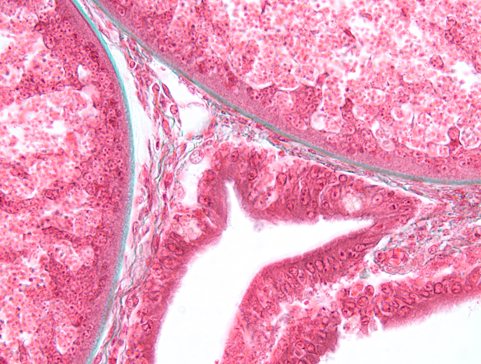
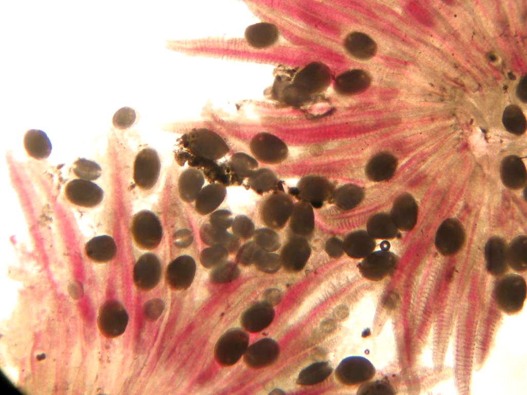
Trematodes (fluk…
Trematodes (fluk…
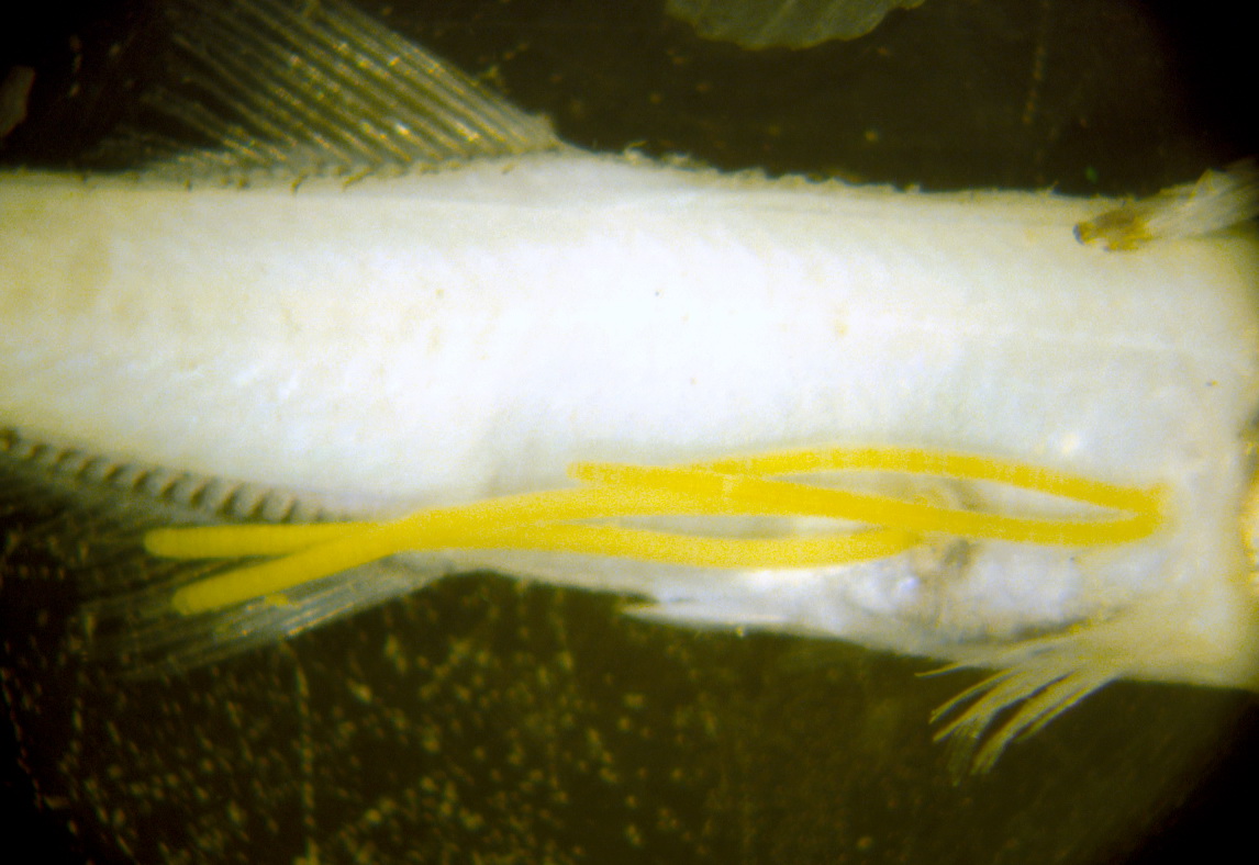
Lernaea (anchor worm…
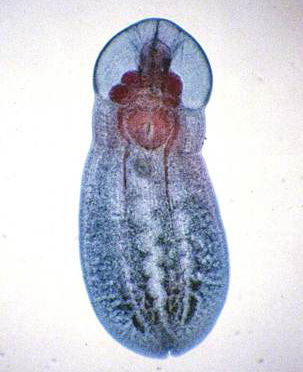
Neascus
Trematodes (fluk…
Myxobolus insidiosu…
Henneguya salminic…
Trematodes (fluk…
…
Clinostomum (Yel…
Glugea
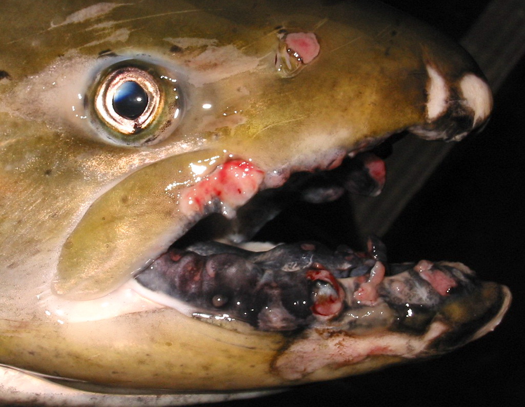
Aeromonas salmonic…
Trematodes (fluk…
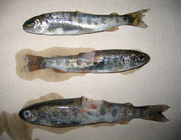
Ceratonova shasta
Nanophyetus
Aca…
EIBS
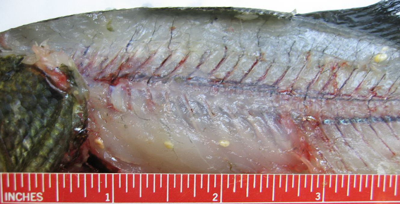
Edwardsiella ictal…
Copepods
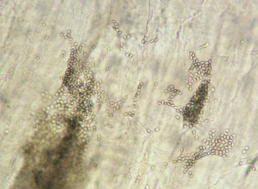
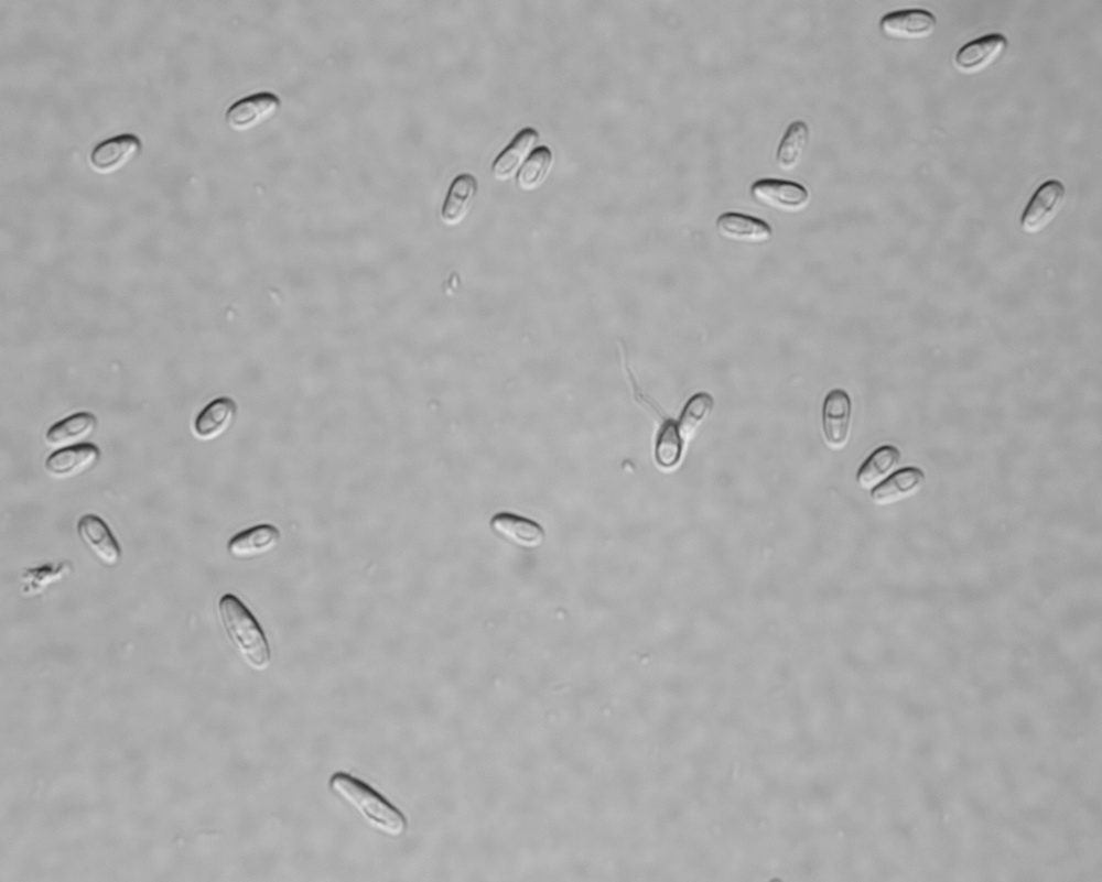
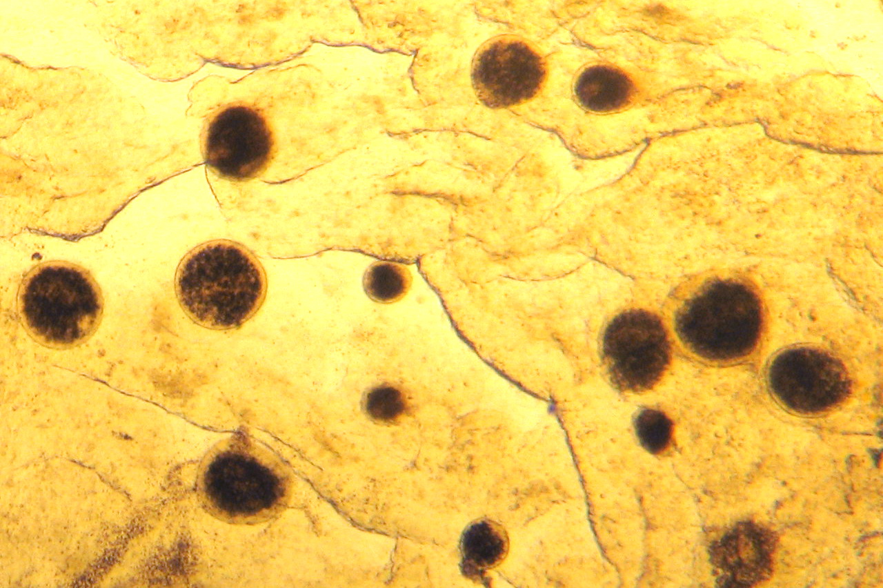
Loma
Myxobolus cerebrali…
Trichophrya
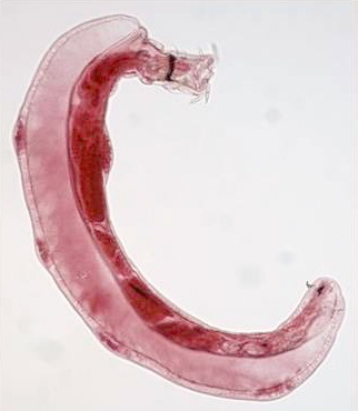
Eustrongylides
Dermocystidium
Ichthyobodo
Henneguya salminic…
Gyrodactylus
…
…
Glugea
Myxobolus cerebrali…
Neoechinorhynchus s…
Mycobacterium mari…
Ichthyophonus
Prolife…
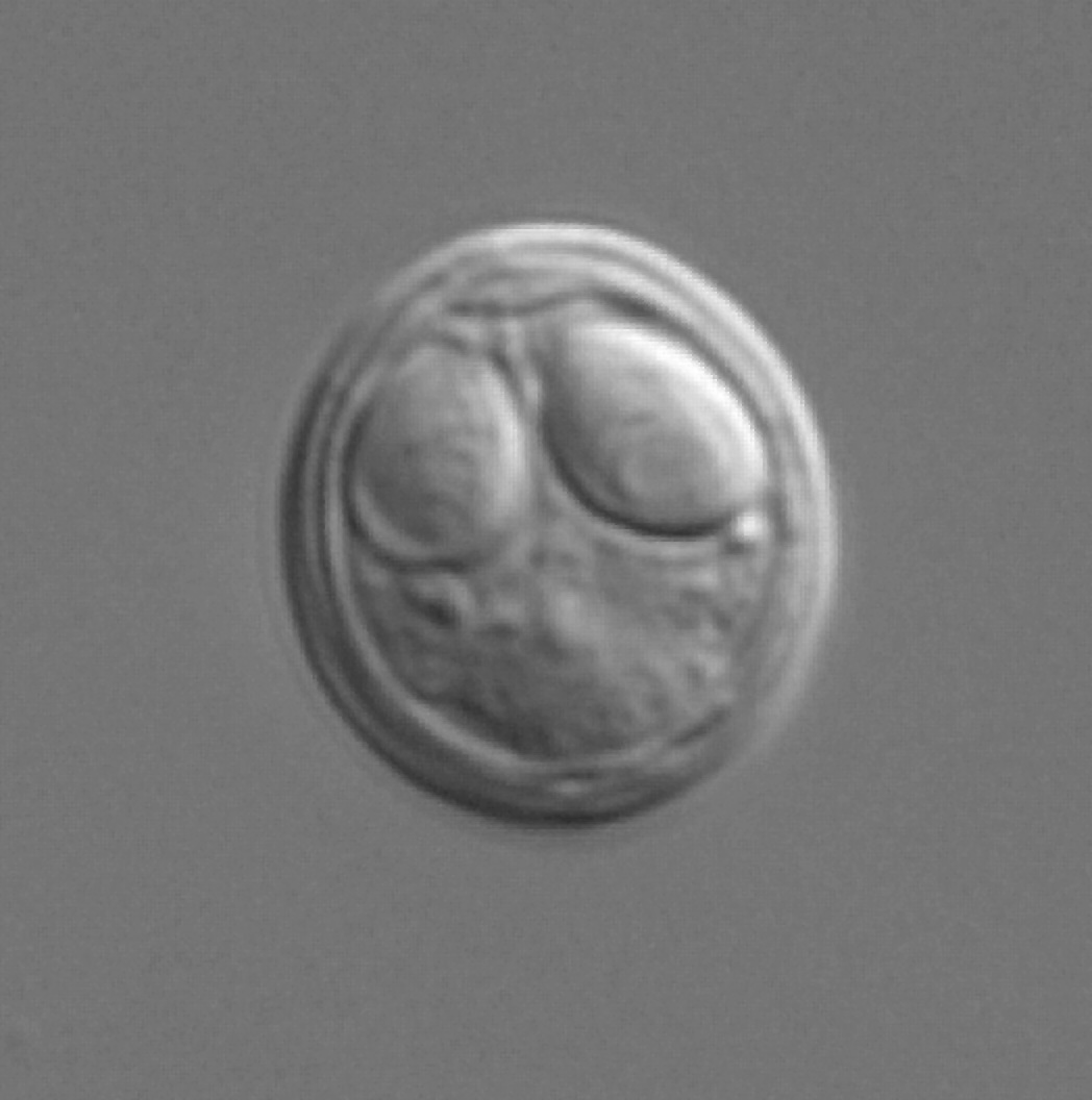
Gloch…
Microsporidia
Fungi and fun…
Aca…
Ceratonova shasta
Gloch…
Posthodiplo…
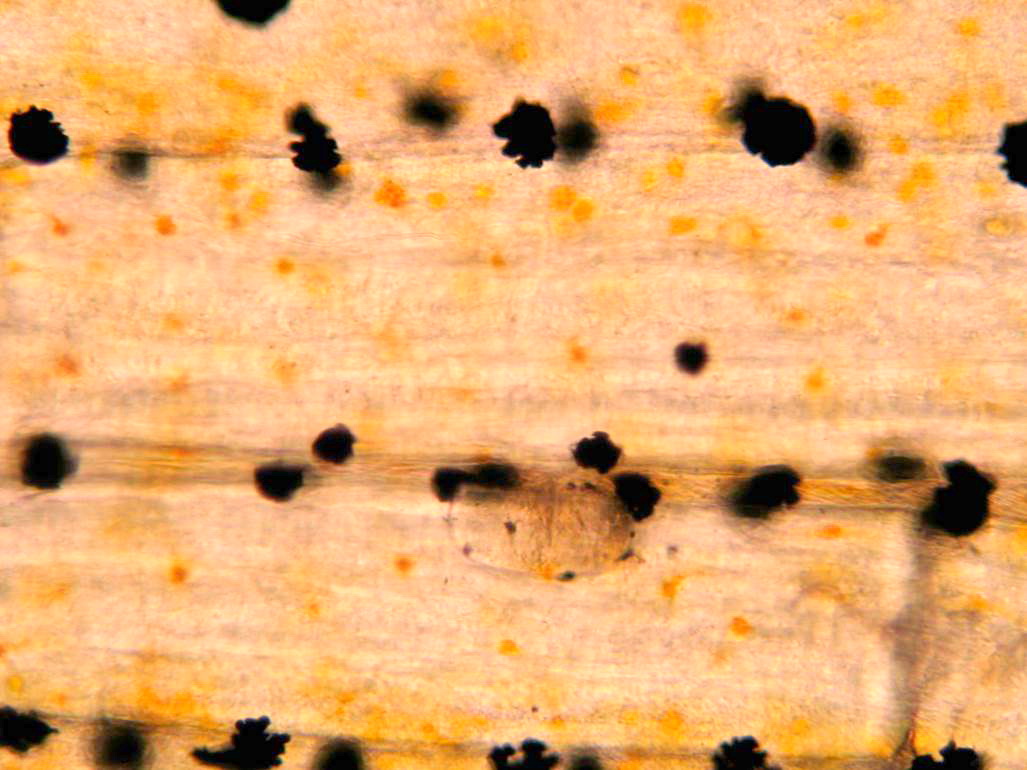
Tumor-like lumps
Ichthyo…
Myxobolus cerebrali…
Pagination
1
Next page
RSS feed