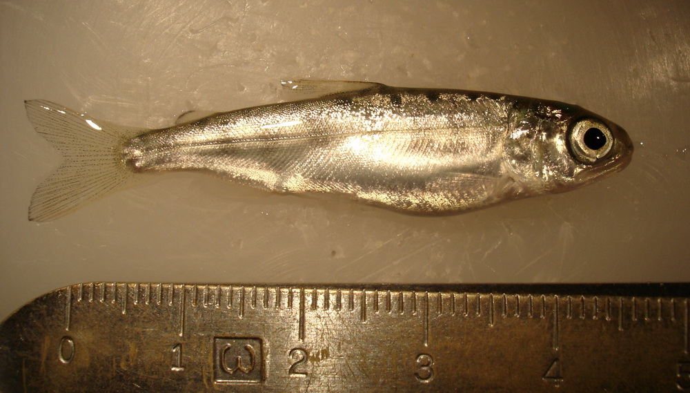
 | DSC00541_RBT.jpg | Fact Sheet Images, Hosts and other organisms | rainbow trout |
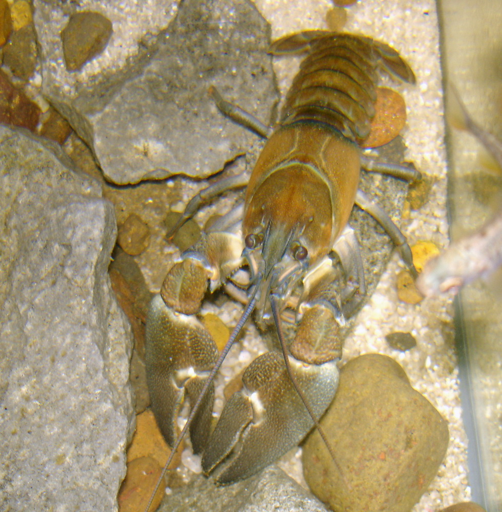
 | DSC00670_crayfish.jpg | Fact Sheet Images, Hosts and other organisms | signal crayfish |
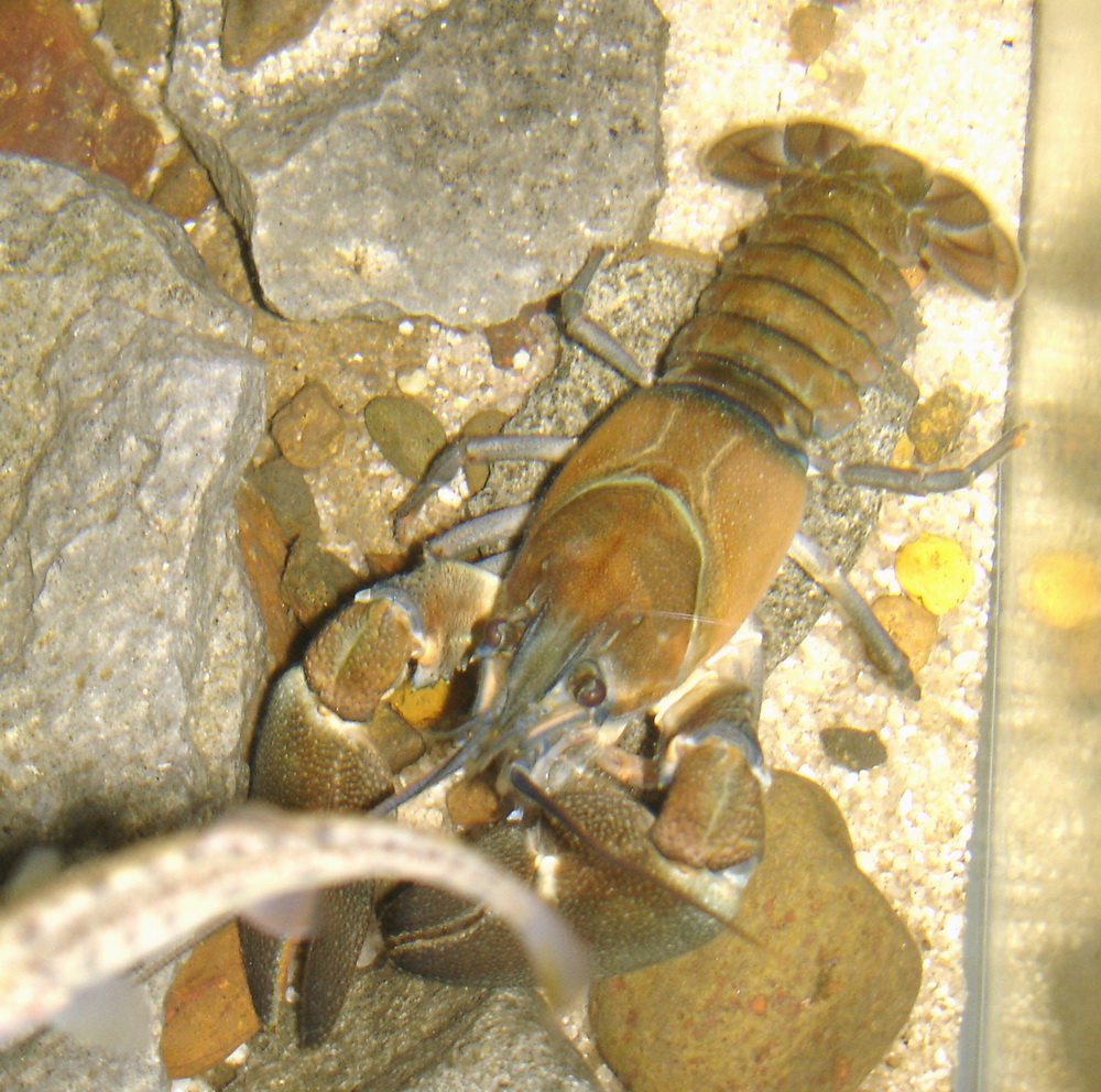
 | DSC00671_crayfish.jpg | Fact Sheet Images, Hosts and other organisms | signal crayfish |
 | DSC01797_RBT.jpg | Fact Sheet Images, Hosts and other organisms | rainbow trout |
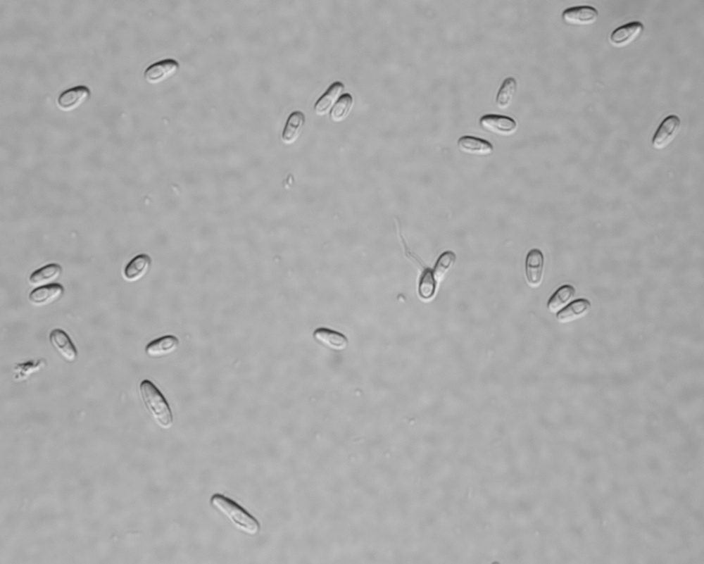
 | SA_Loma_01a_x10bf_9shot.jpg | All Pathogens, Fact Sheet Images | |
 | SA_Loma_02b_x40bf_4shot.jpg | All Pathogens, Fact Sheet Images | |
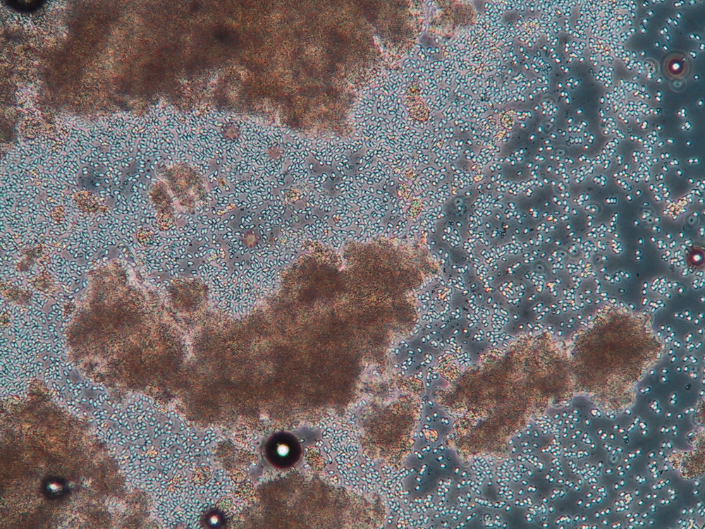
 | SA_other_para_01_x20nom_4shot_coho_kidney.jpg | All Pathogens, Fact Sheet Images | coho salmon |
 | SA_other_para_02_x20nom_4shot_coho_kidney.jpg | All Pathogens, Fact Sheet Images | coho salmon |
 | SA_other_para_03_x20nom_4shot_coho_kidney.jpg | All Pathogens, Fact Sheet Images | coho salmon |
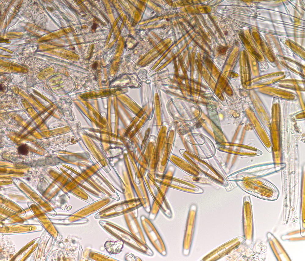
 | SA_alga01_x40bf_9shot.jpg | Fact Sheet Images, Hosts and other organisms | algae |
 | SA_alga02_x40bf_9shot.jpg | Fact Sheet Images, Hosts and other organisms | algae |
 | SA_alga03_x40bf_9shot.jpg | Fact Sheet Images, Hosts and other organisms | algae |
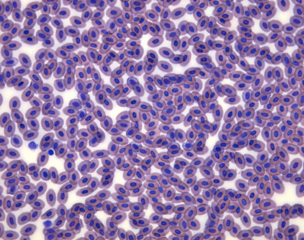
 | SA_blood_smear_20061013B_01a_x63bf_9_DQ.jpg | Fact Sheet Images, Hosts and other organisms | rainbow trout |
 | SA_DSC00990.jpg | Fact Sheet Images, Hosts and other organisms | rainbow trout |
 | SA_DSC02921.jpg | All Pathogens, Fact Sheet Images | Tubifex |
 | SA_DSC04356ed.jpg | All Pathogens, Fact Sheet Images | |
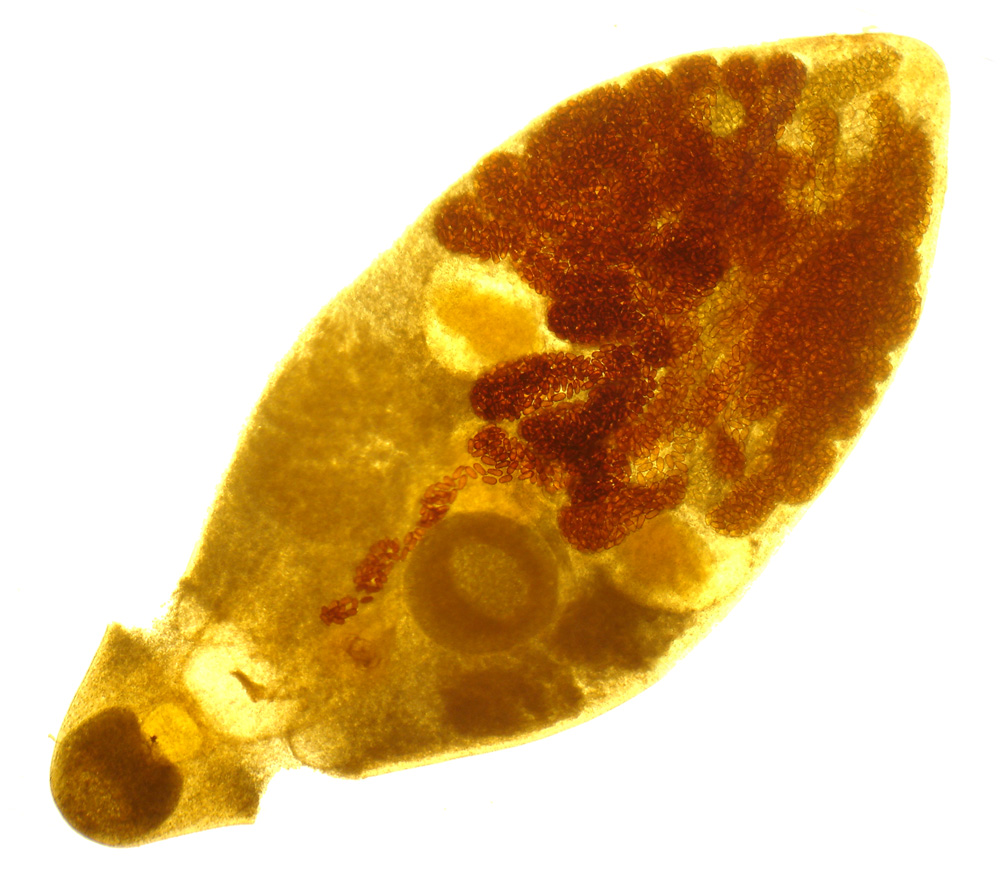
 | SA_DSC05503.jpg | Fact Sheet Images, Hosts and other organisms | bryozoan |
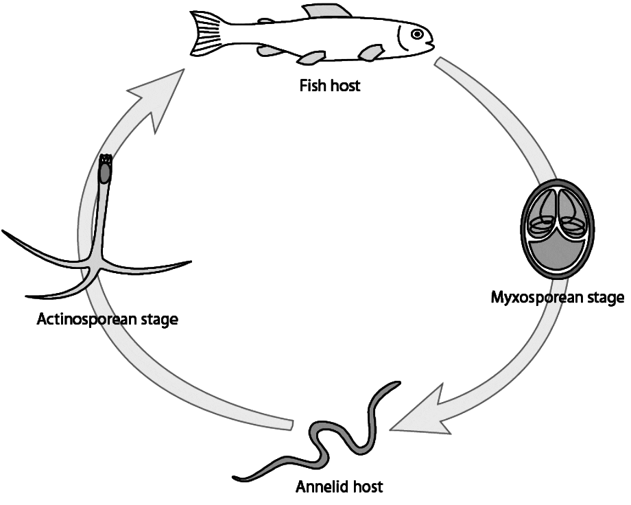
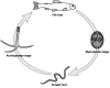 | generic_lifecycle.gif | Illustration - life cycle, Fact Sheet Images | |
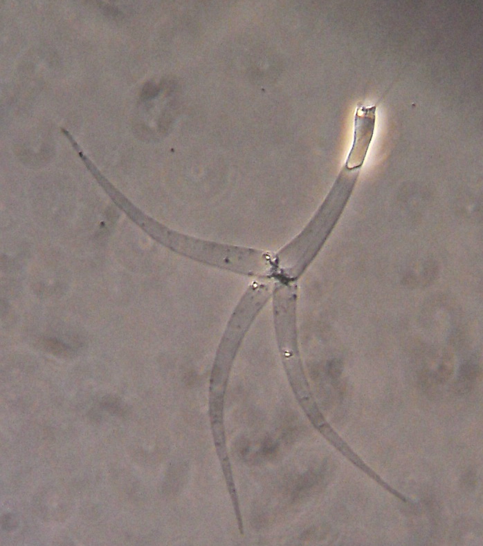
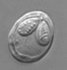
 | SA_Generic_myxobolus.jpg | All Pathogens, Fact Sheet Images | |
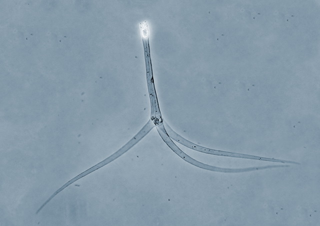
 | SA_Generic_TAM.jpg | All Pathogens, Fact Sheet Images | |
 | SA_IMG_4724_crop.jpg | All Pathogens, Fact Sheet Images | common carp |
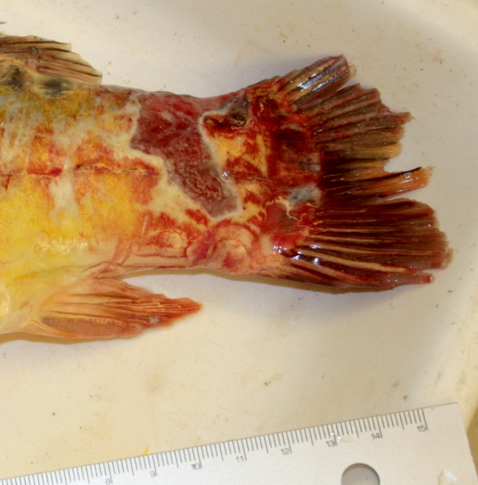
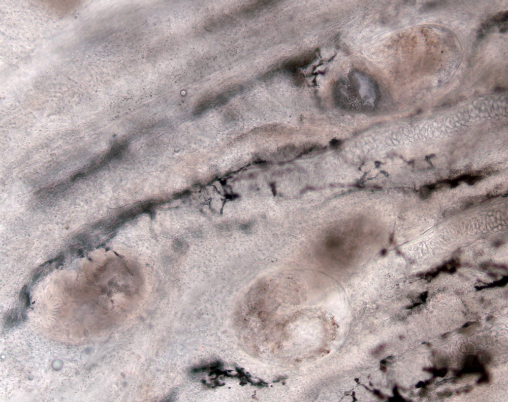
 | SA_157_5708.jpg | Hosts and other organisms, Fact Sheet Images | sculpin (unspecified) |
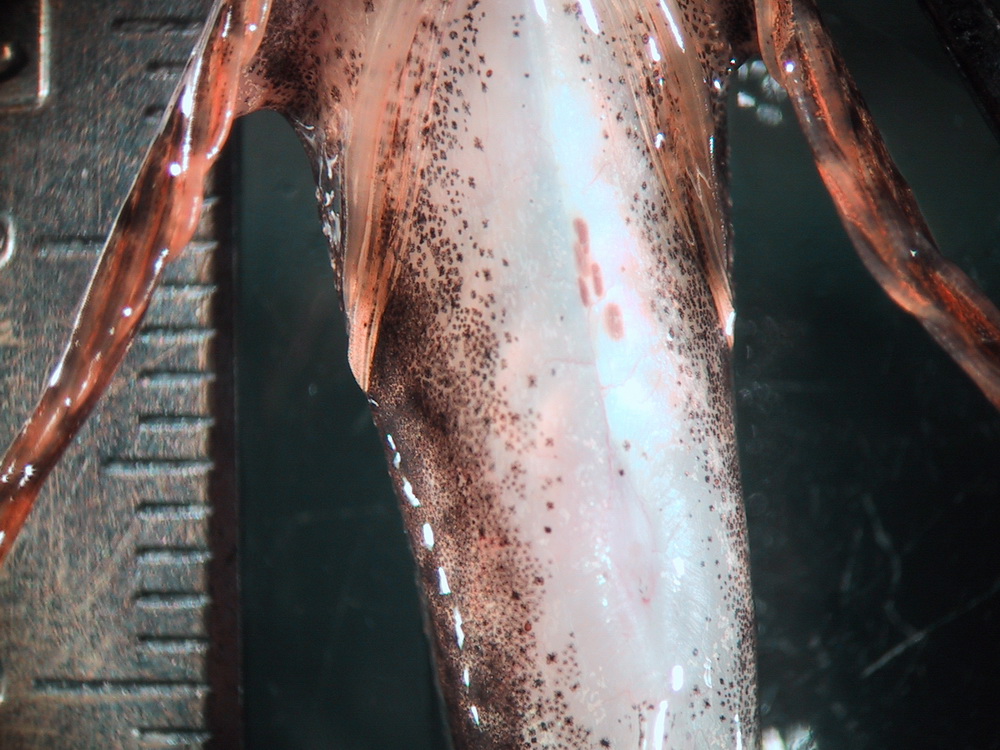
 | SA_157_5709.jpg | All Pathogens, Fact Sheet Images | sculpin (unspecified) |
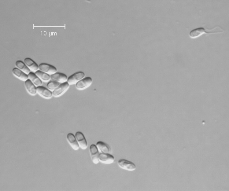
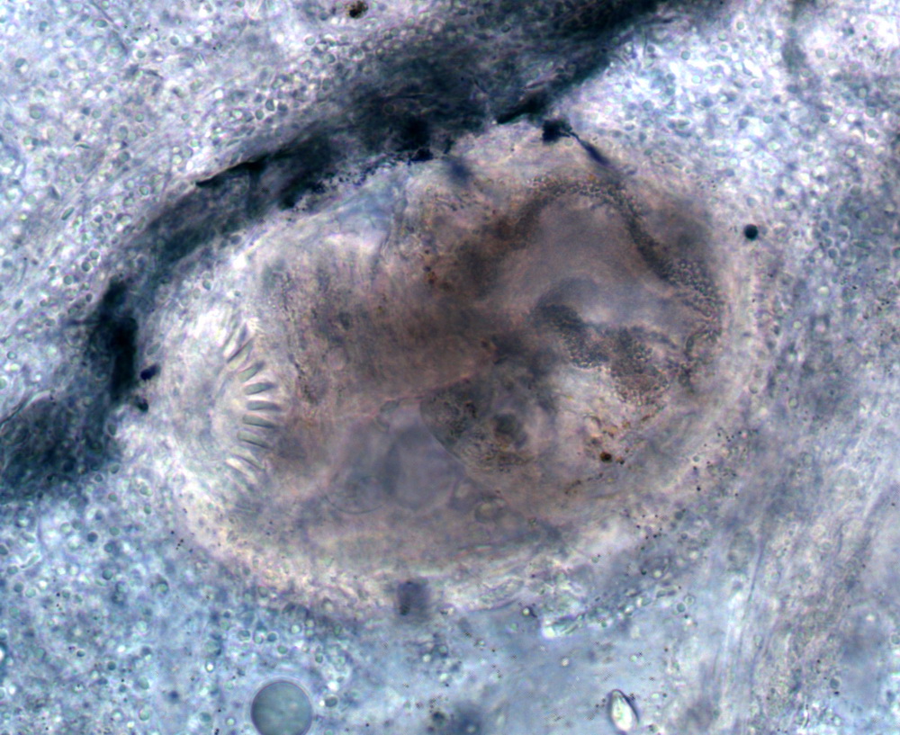
 | SA_MYX_CC02calib.jpg | All Pathogens, Fact Sheet Images | sculpin (unspecified) |
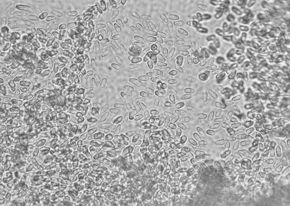
 | SA_MYXCC02_01_x40.jpg | All Pathogens, Fact Sheet Images | sculpin (unspecified) |
 | SA_MYXCC02_09_x100.jpg | All Pathogens, Fact Sheet Images | sculpin (unspecified) |
 | SA_MYXCC02_02_x100.jpg | All Pathogens, Fact Sheet Images | sculpin (unspecified) |
 | SA_157_5710.jpg | All Pathogens, Fact Sheet Images | sculpin (unspecified) |
 | SA_dactylo_20120914_01a_x20bf9h.jpg | All Pathogens, Fact Sheet Images | Oregon chub |
 | SA_dactylo_20120914_01b_x40bf9h.jpg | All Pathogens, Fact Sheet Images | Oregon chub |