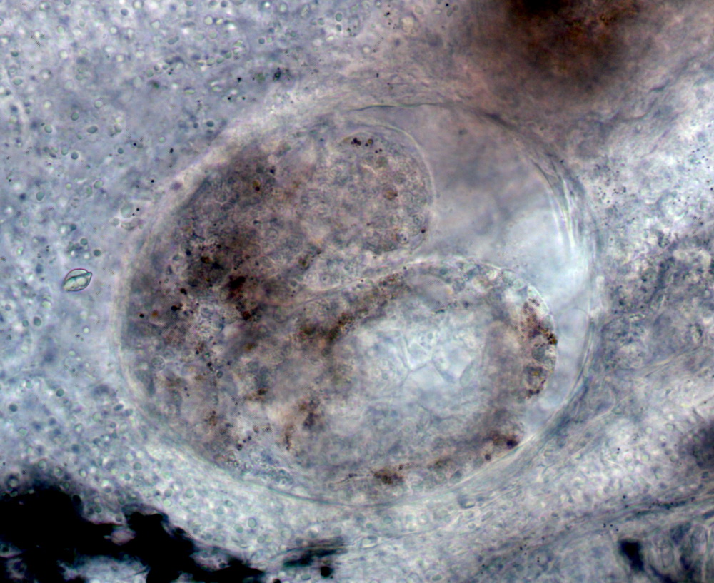
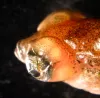
 | SA_dactylo_20120914_01c_x40bf9h.jpg | All Pathogens, Fact Sheet Images | Oregon chub |
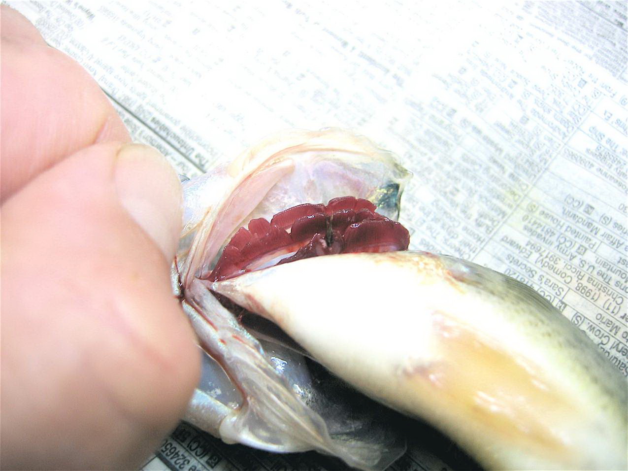
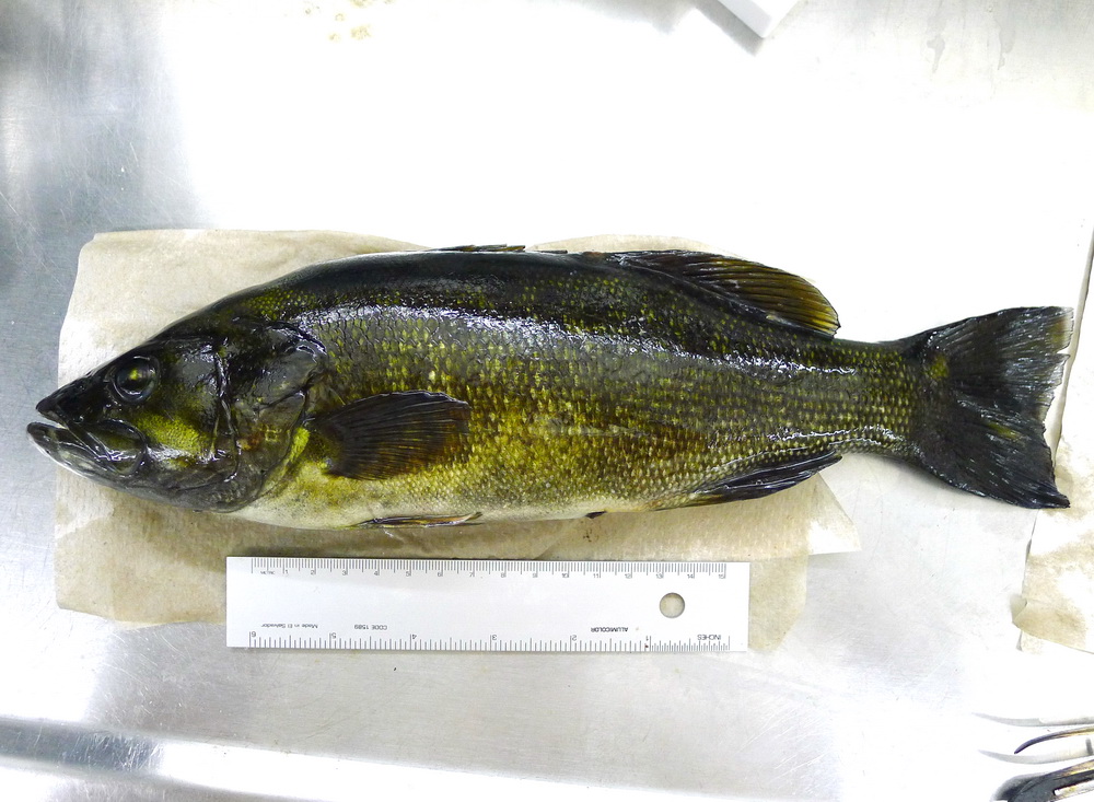
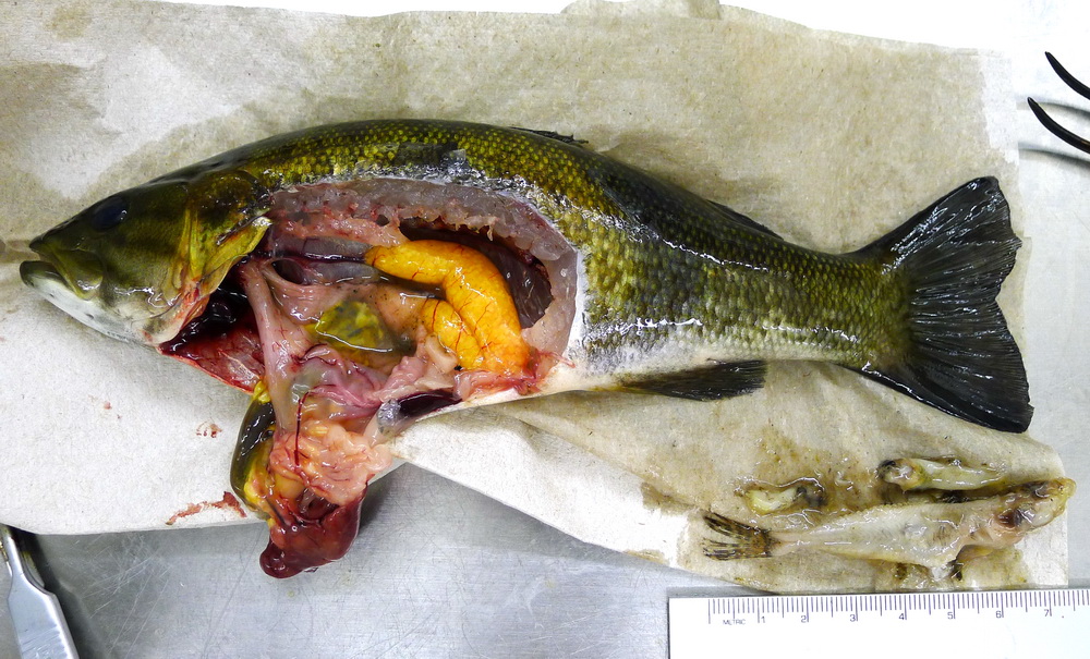
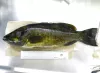
 | seedyLMB_CB01.jpg | All Pathogens, Fact Sheet Images | largemouth bass |
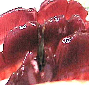
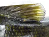
 | seedyLMB_CB02.jpg | All Pathogens, Fact Sheet Images | largemouth bass |
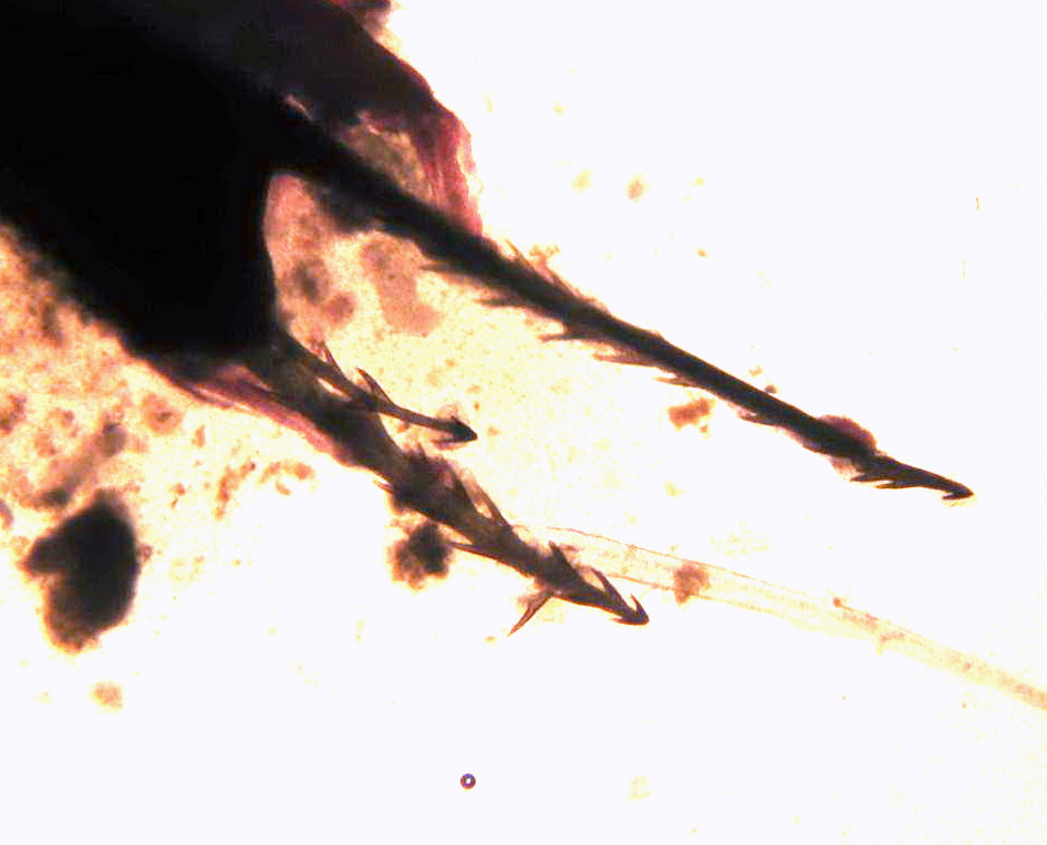
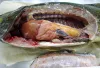
 | seedyLMB_CB03.jpg | All Pathogens, Fact Sheet Images | largemouth bass |
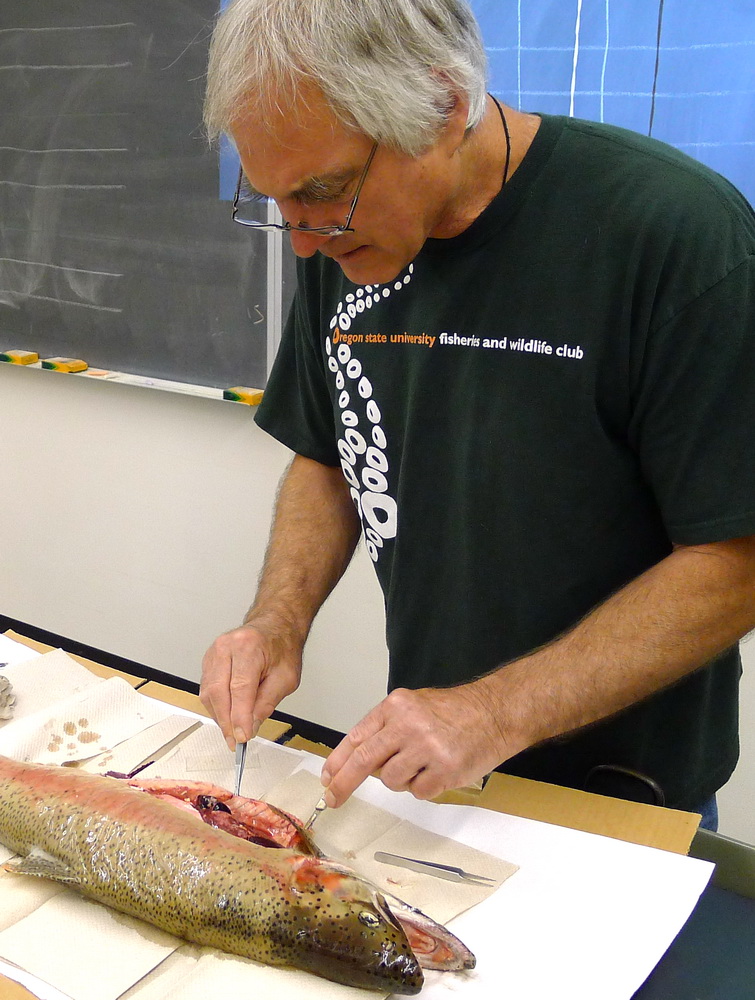
 | SA_Banner_P1050450.jpg | Personel, Fact Sheet Images | |
 | SA_P1020178_yellowgrub.jpg | All Pathogens, Fact Sheet Images | smallmouth bass |
 | P1020173_SA_yperch.jpg | Fact Sheet Images, Hosts and other organisms | yellow perch |
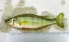
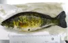
 | P1020174_SA_smb.jpg | Hosts and other organisms, Fact Sheet Images | smallmouth bass |
 | P1020175_SA_smb.jpg | All Pathogens, Fact Sheet Images | smallmouth bass |
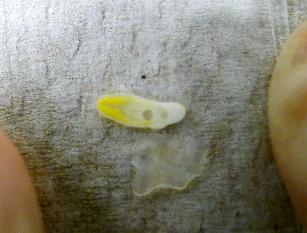
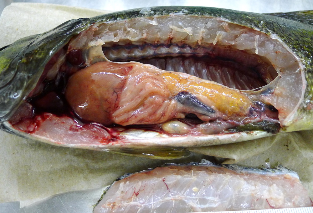
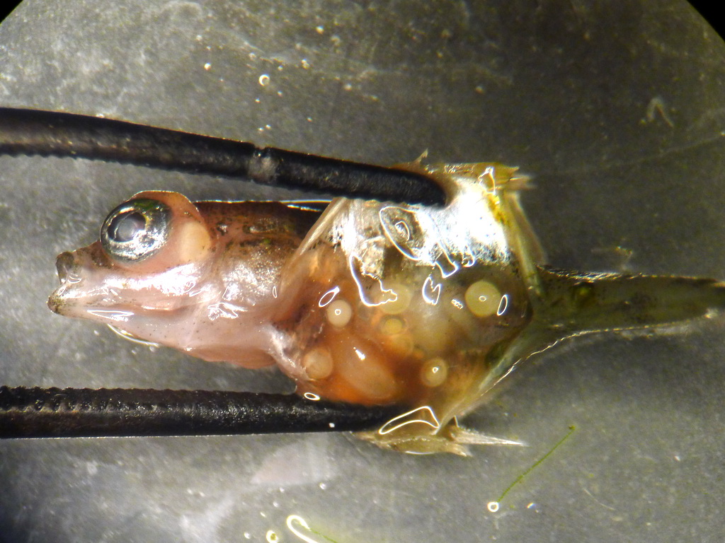
 | P1020176_SA_yellowgrub.jpg | All Pathogens, Fact Sheet Images | smallmouth bass |
 | P1020177_SA_smb.jpg | Fact Sheet Images, Hosts and other organisms | smallmouth bass |
 | P1020179_SA_smb.jpg | Fact Sheet Images, Hosts and other organisms | smallmouth bass |
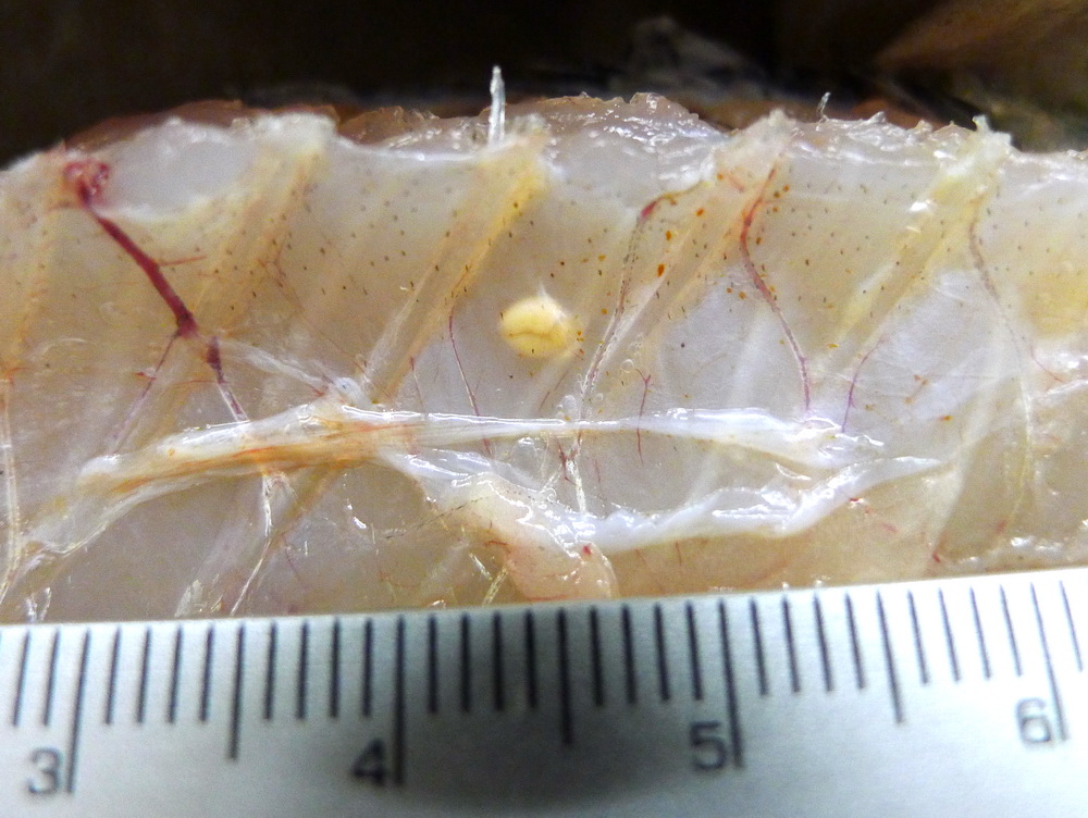
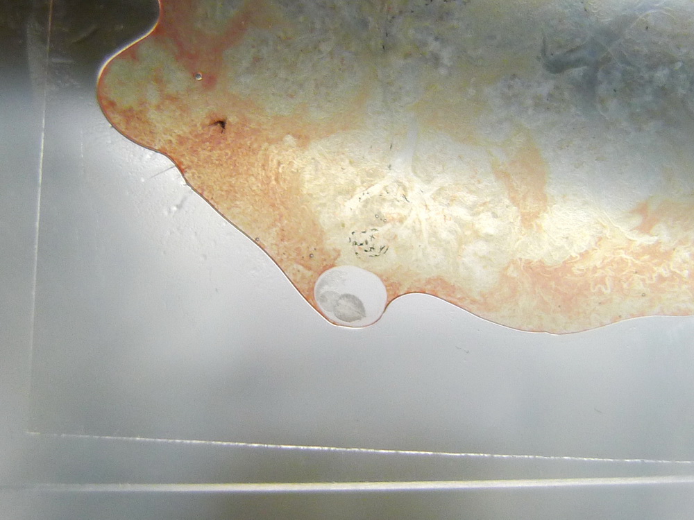
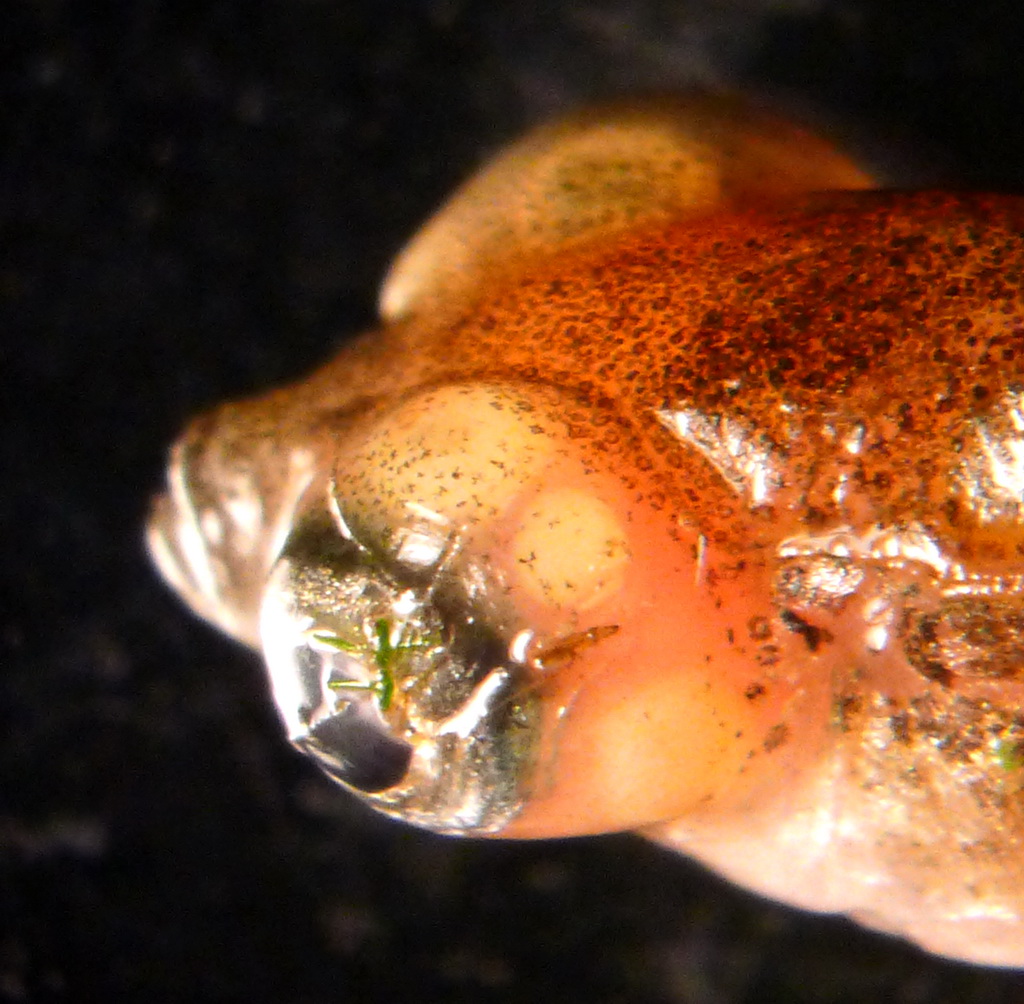
 | P1020180_SA_whitegrub.jpg | All Pathogens, Fact Sheet Images | smallmouth bass |
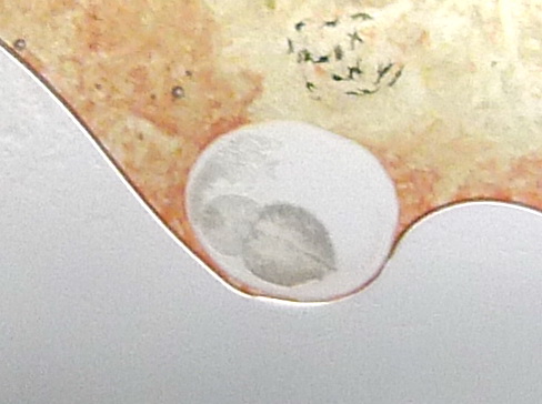
 | P1020180_SA_zoomed.jpg | All Pathogens, Fact Sheet Images | smallmouth bass |
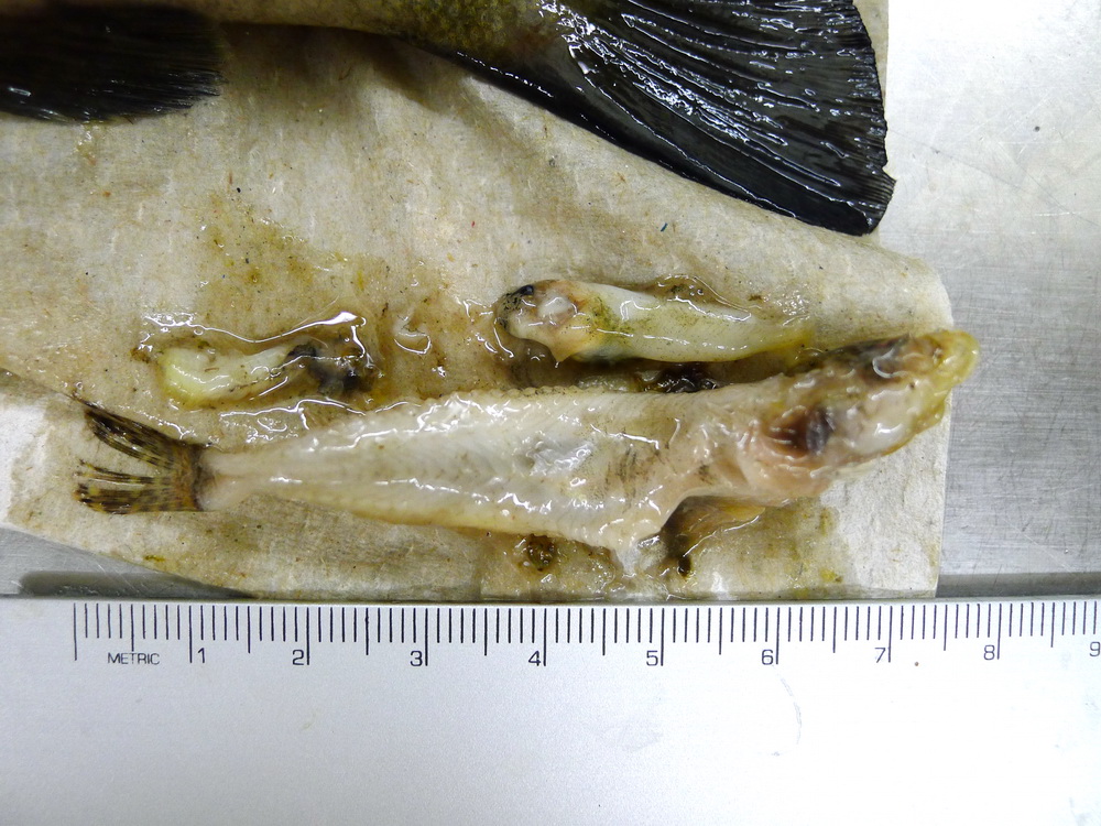
 | P1020182_SA_stomachcontent.jpg | Fact Sheet Images, Hosts and other organisms | smallmouth bass |
 | P1020183_SA_smb.jpg | Fact Sheet Images, Hosts and other organisms | smallmouth bass |
 | Myxobilatus_11M21.jpg | All Pathogens, Fact Sheet Images | largemouth bass |
 | 11M21_x100nm9h_04a.jpg | All Pathogens, Fact Sheet Images | largemouth bass |
 | P1020181_SA.jpg | All Pathogens, Fact Sheet Images | smallmouth bass |
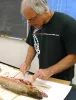
 | P1050695_sampling_CogswellCk.jpg | Field Localities (-> and enter Locality below), Personel, Fact Sheet Images | |
 | P1060313_Cshasta.jpg | All Pathogens, Fact Sheet Images | rainbow trout |
 | P1060316_Cshasta.jpg | All Pathogens, Fact Sheet Images | rainbow trout |
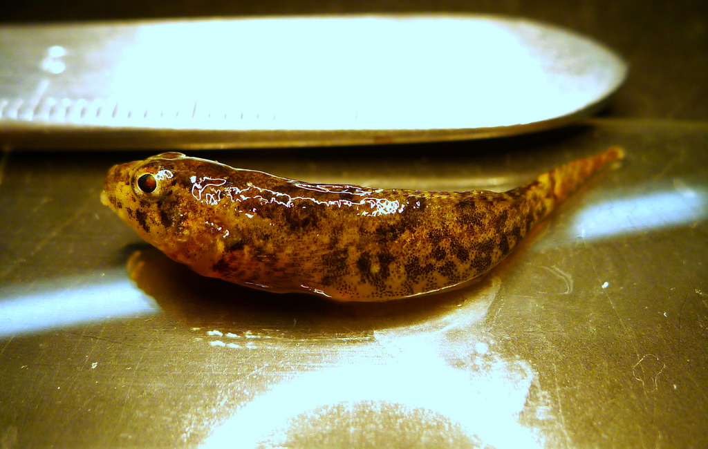
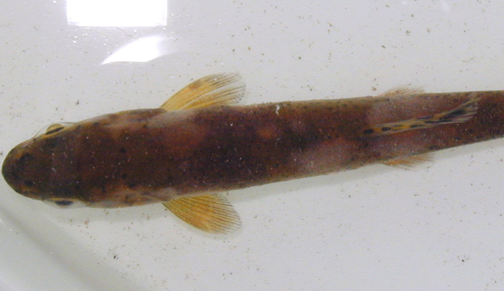
 | P1060632_sculpin.jpg | Fact Sheet Images, Hosts and other organisms | sculpin (unspecified) |
 | P1060629_diplostomum.jpg | All Pathogens, Fact Sheet Images | threespine stickleback |
 | P1060631_diplostomum.jpg | All Pathogens, Fact Sheet Images | threespine stickleback |
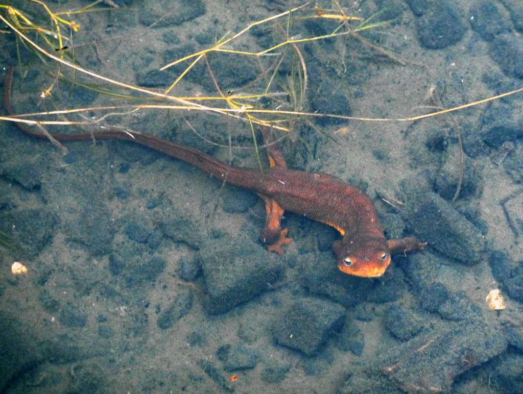
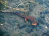 | P1000836_newt.jpg | Hosts and other organisms, Fact Sheet Images | rough-skinned newt; salamander |
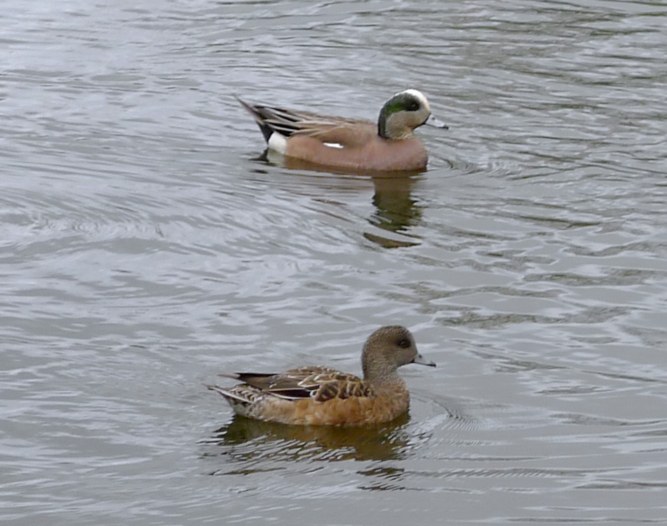
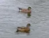 | P1080719_widgeon.jpg | Hosts and other organisms, Fact Sheet Images | American wigeon |
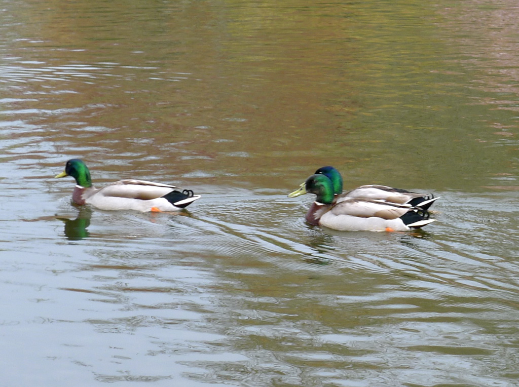
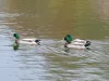 | P1080717_mallards.jpg | Hosts and other organisms, Fact Sheet Images | mallard duck |
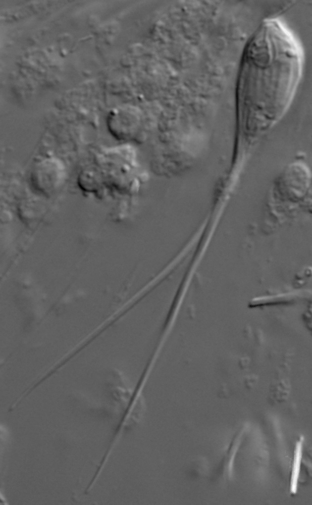
 | CB_Apiostoma_siletz_Good2.jpg | All Pathogens, Fact Sheet Images | |
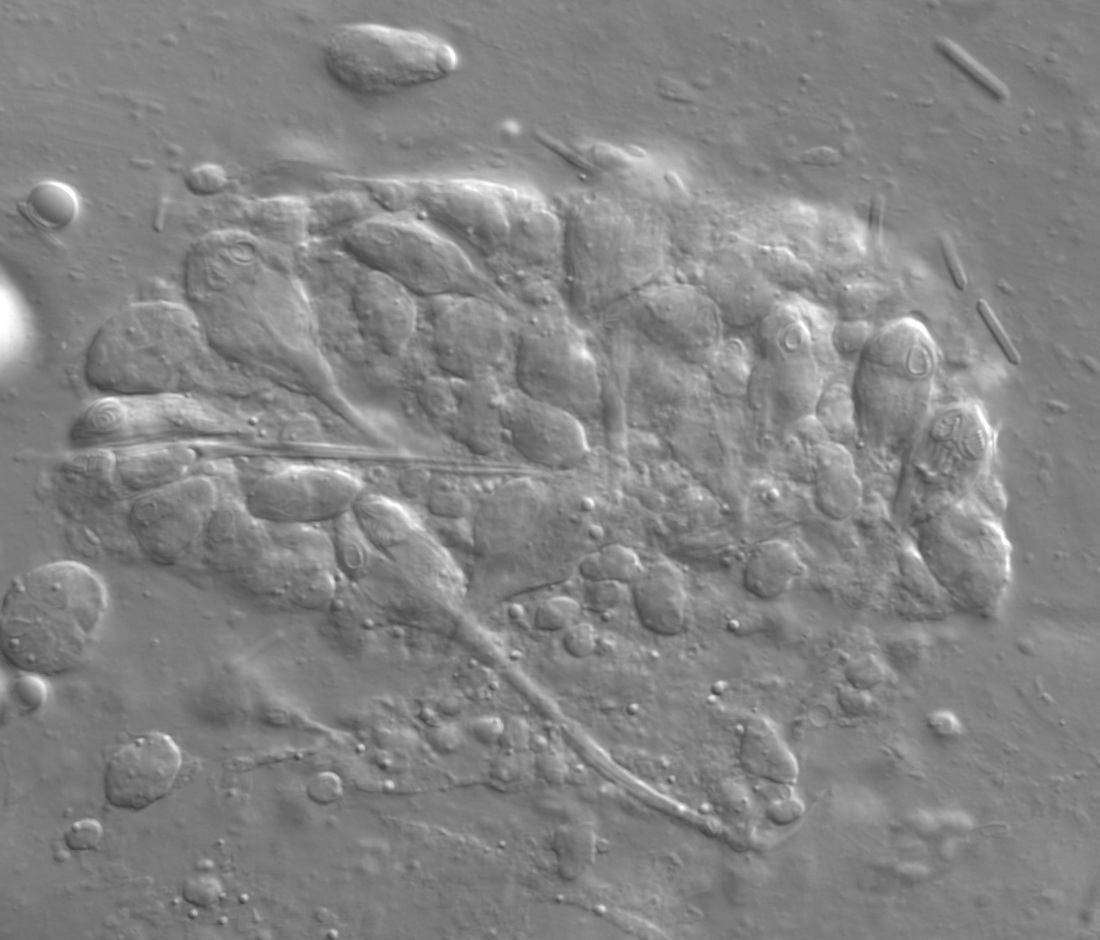
 | CB_Apiostoma_siletz_Good3.jpg | All Pathogens, Fact Sheet Images | |