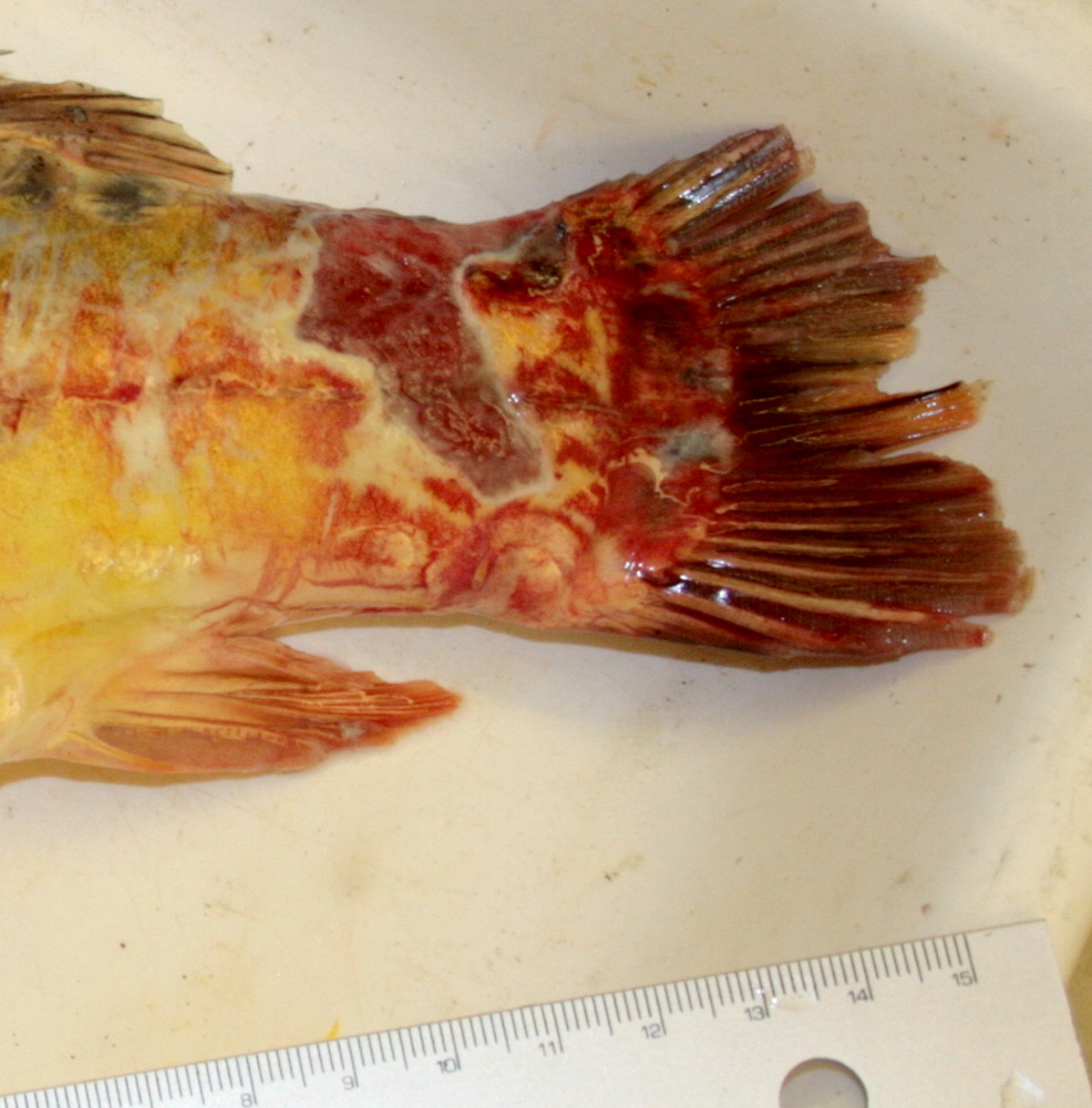
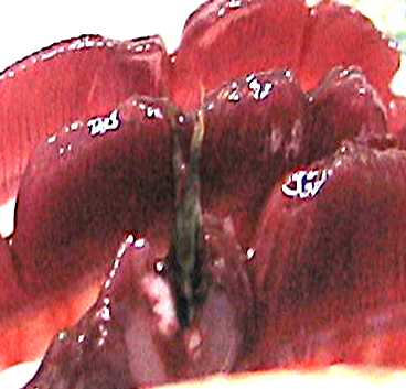
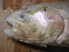
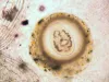
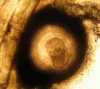
 | SA_157_5709.jpg | All Pathogens, Fact Sheet Images | sculpin (unspecified) |
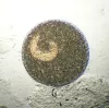
 | seedyLMB_CB01.jpg | All Pathogens, Fact Sheet Images | largemouth bass |
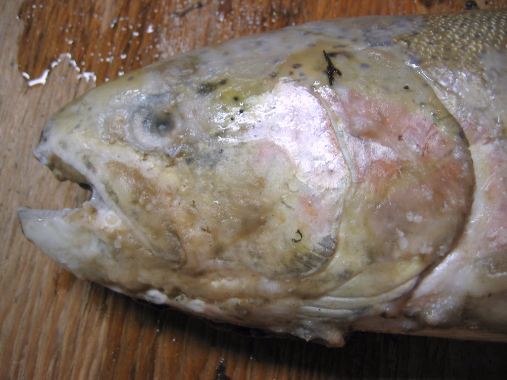
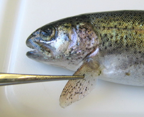
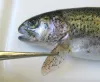
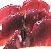
 | CB_StW_fungus2.jpg | All Pathogens, Fact Sheet Images | Winter steelhead trout |
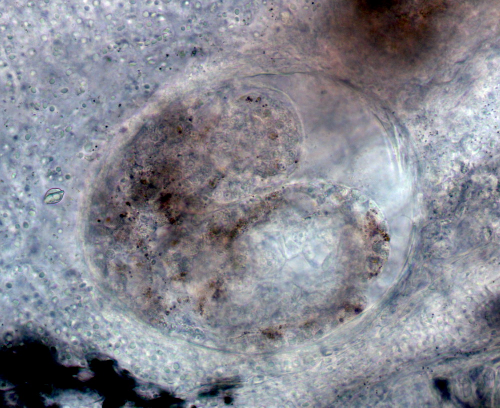
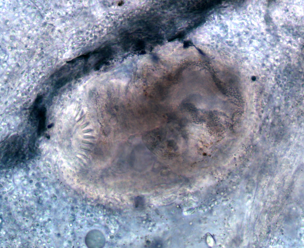
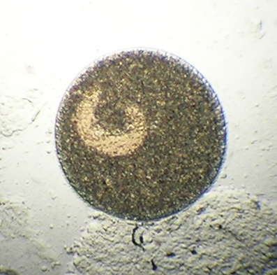
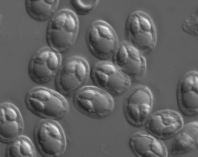
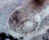 | SA_dactylo_20120914_01c_x40bf9h.jpg | All Pathogens, Fact Sheet Images | Oregon chub |
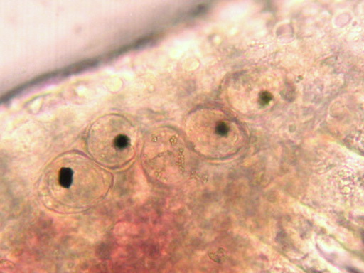
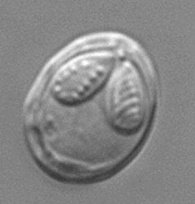
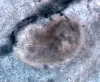 | SA_dactylo_20120914_01b_x40bf9h.jpg | All Pathogens, Fact Sheet Images | Oregon chub |
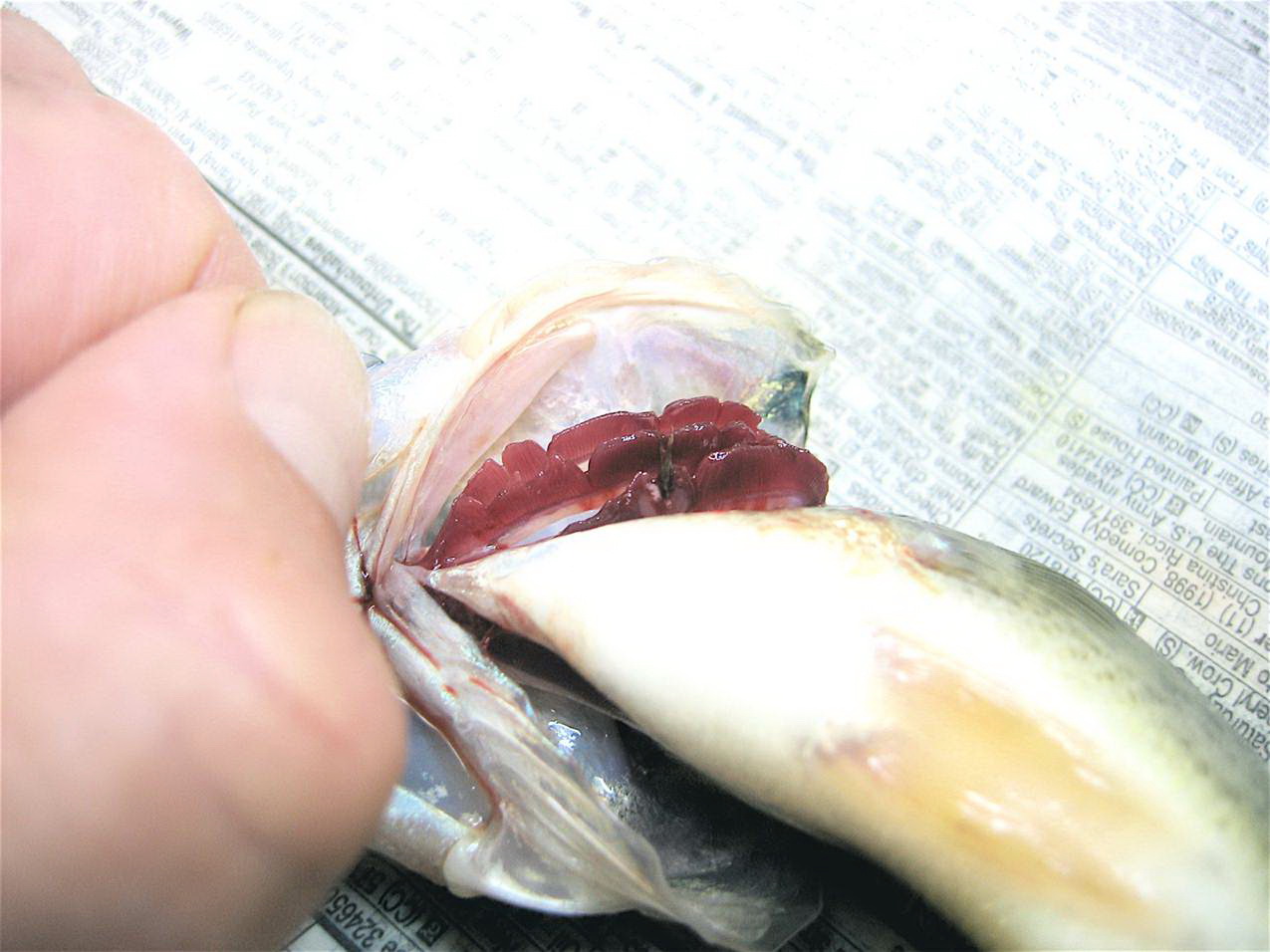
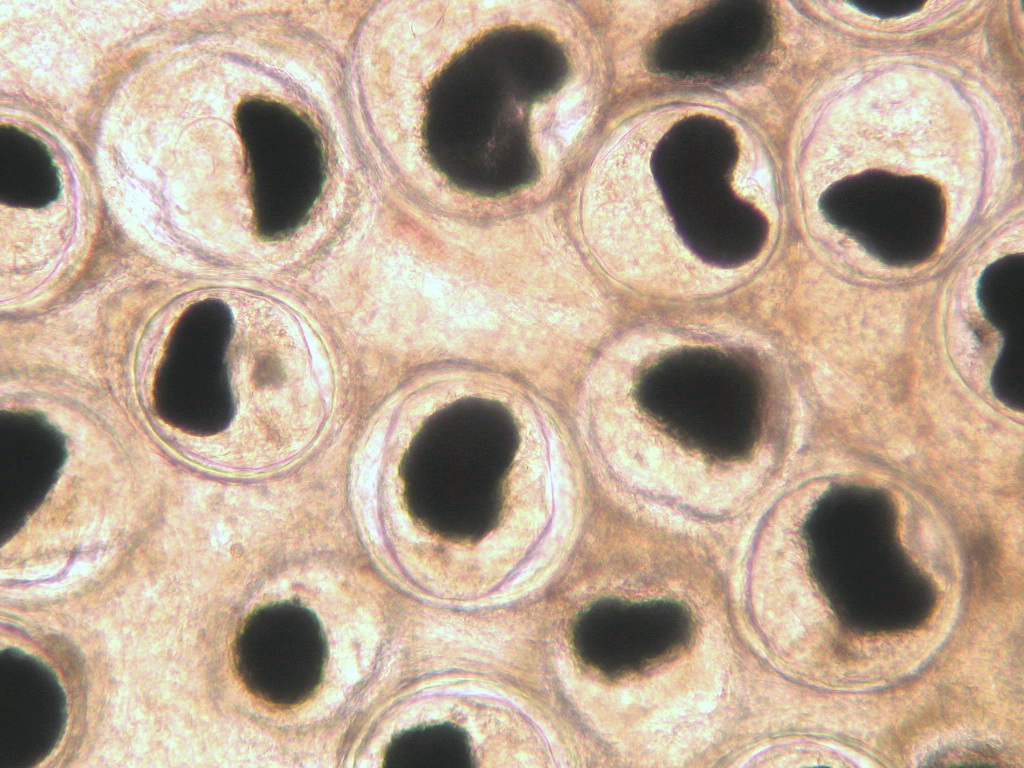
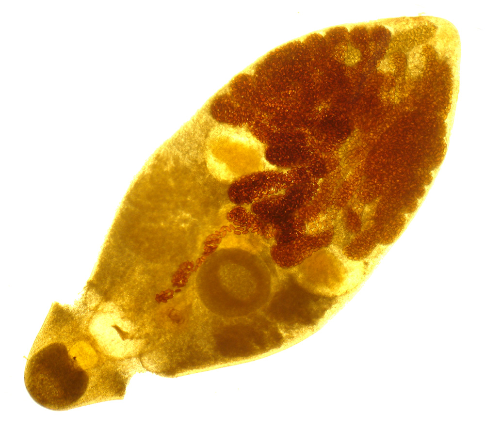
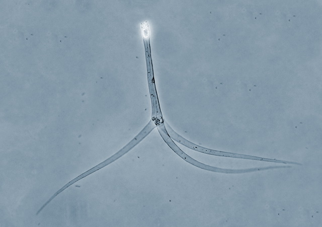
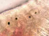 | CB_Sanguin8.jpg | All Pathogens, Fact Sheet Images | |
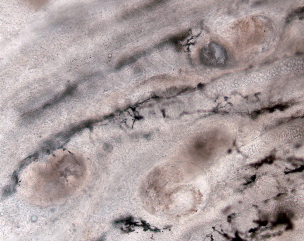
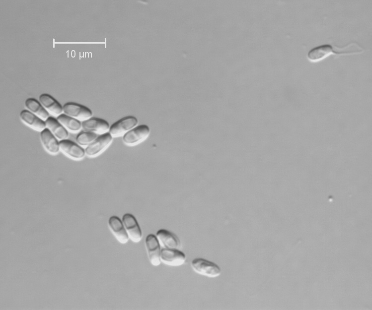
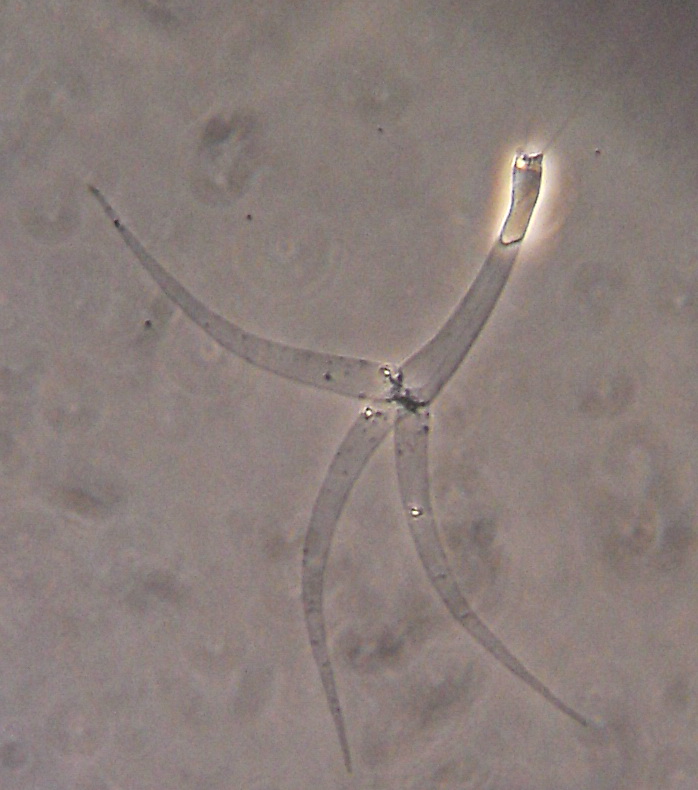
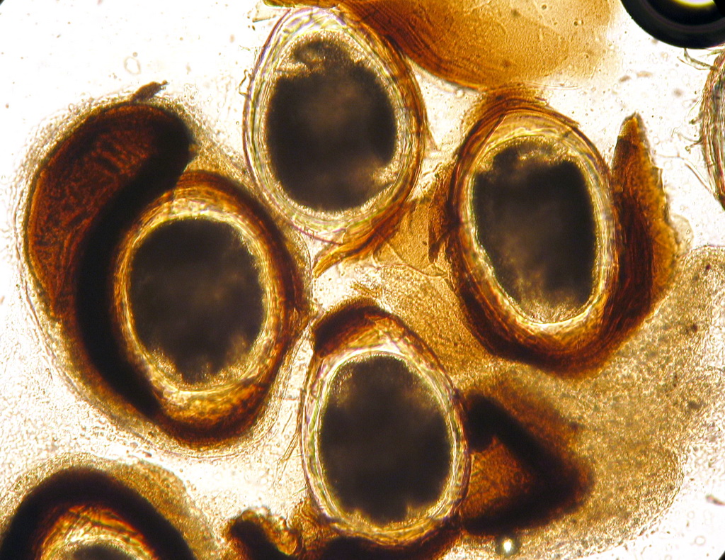
 | SA_dactylo_20120914_01a_x20bf9h.jpg | All Pathogens, Fact Sheet Images | Oregon chub |
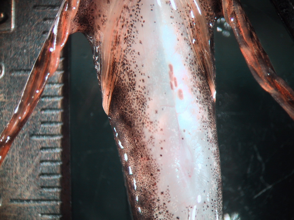
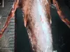
 | SA_157_5710.jpg | All Pathogens, Fact Sheet Images | sculpin (unspecified) |
 | CB_PGE_Fung1.jpg | All Pathogens, Fact Sheet Images | |
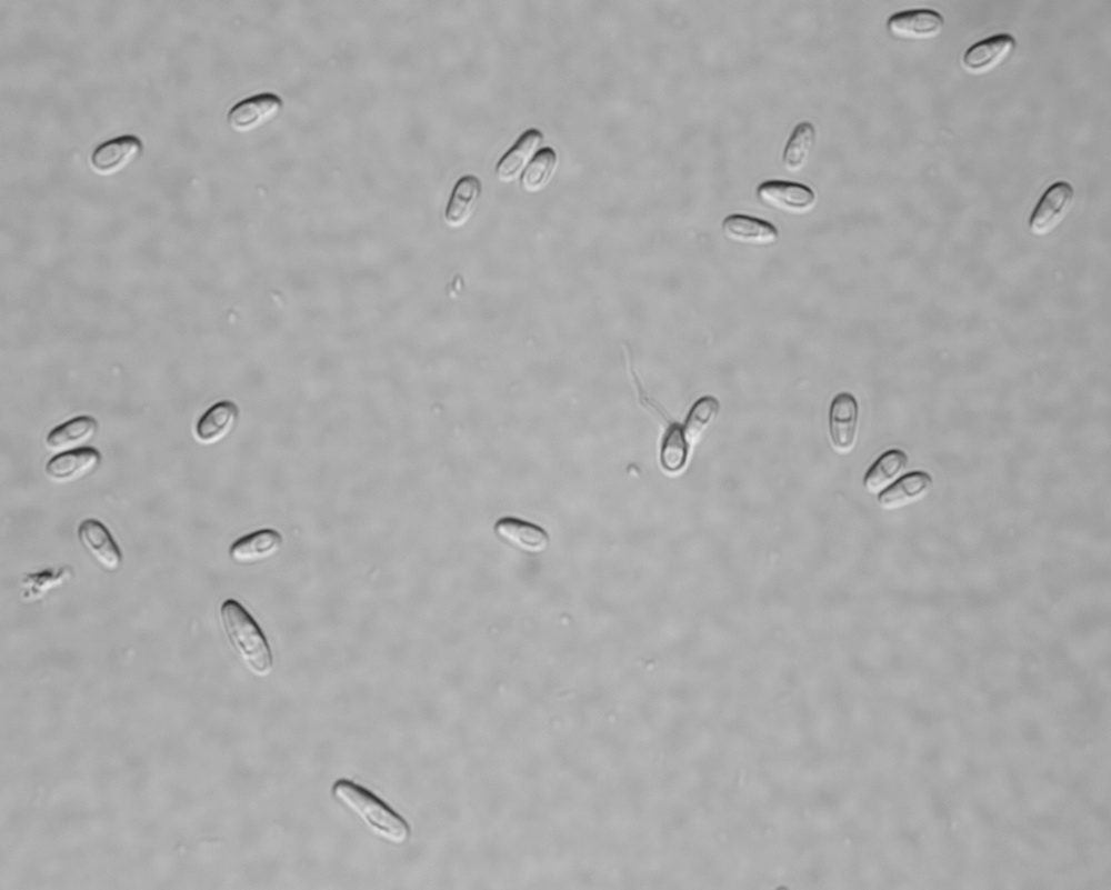
 | SA_MYXCC02_02_x100.jpg | All Pathogens, Fact Sheet Images | sculpin (unspecified) |
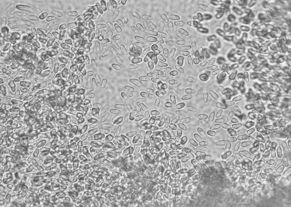
 | SA_MYXCC02_09_x100.jpg | All Pathogens, Fact Sheet Images | sculpin (unspecified) |
 | CB_Nano_muscle7.jpg | All Pathogens, Fact Sheet Images | |
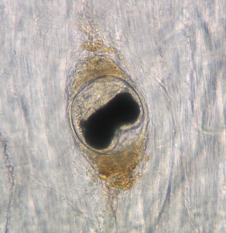
 | SA_MYXCC02_01_x40.jpg | All Pathogens, Fact Sheet Images | sculpin (unspecified) |
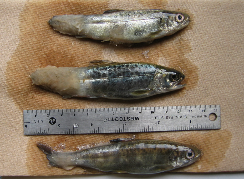
 | SA_MYX_CC02calib.jpg | All Pathogens, Fact Sheet Images | sculpin (unspecified) |
 | CB_Nano_intest7.jpg | All Pathogens, Fact Sheet Images | |
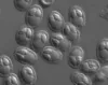
 | SA_Generic_myxobolus.jpg | All Pathogens, Fact Sheet Images | |
 | CB_CohoBlk_spt4.jpg | All Pathogens, Fact Sheet Images | coho salmon |
 | SA_DSC02921.jpg | All Pathogens, Fact Sheet Images | Tubifex |
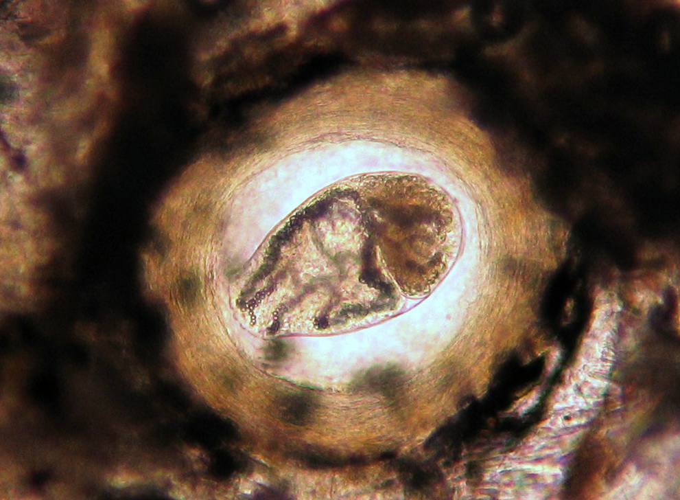
 | SA_DSC04356ed.jpg | All Pathogens, Fact Sheet Images | |
 | CB_CoBLkSpt30.jpg | All Pathogens, Fact Sheet Images | coho salmon |
 | CB_BLK_SPT1.jpg | All Pathogens, Fact Sheet Images | rainbow trout |
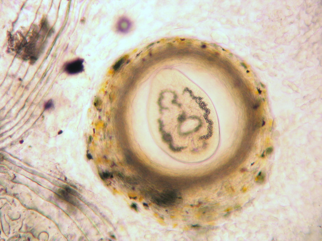
 | SA_DSCN0583.jpg | All Pathogens | |
 | CB_BLk_SPOT2.jpg | All Pathogens, Fact Sheet Images | rainbow trout |
 | SA_IMG_4724.jpg | All Pathogens, Hosts and other organisms | common carp |
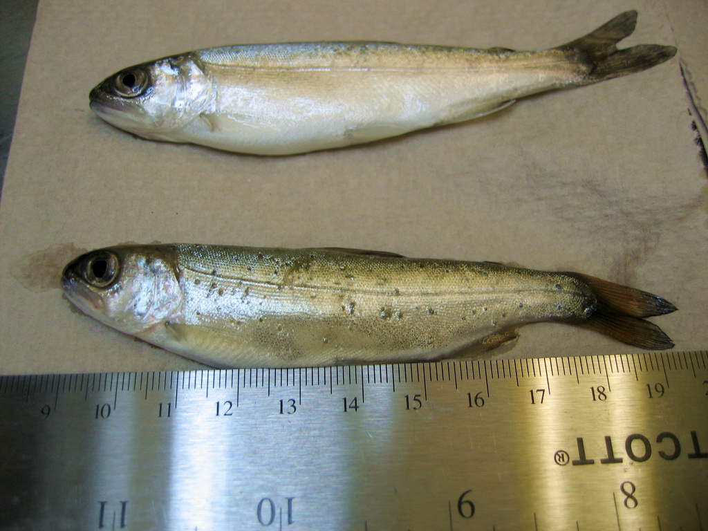
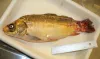
 | CB_146_4665.jpg | All Pathogens, Fact Sheet Images | threespine stickleback |
 | SA_IMG_4724_crop.jpg | All Pathogens, Fact Sheet Images | common carp |
 | CB_BlacSptSucker.jpg | All Pathogens, Fact Sheet Images | sucker (unidentified) |
 | seedyLMB_CB02.jpg | All Pathogens, Fact Sheet Images | largemouth bass |
 | 20120911e_07a_x100nm1h.jpg | All Pathogens, Fact Sheet Images | redside shiner |
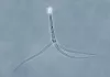 | SA_Generic_TAM.jpg | All Pathogens, Fact Sheet Images | |