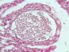
 | Slide # 42 Kidney Flavobacterium psychrophilus Steiner 400X.jpg | All Pathogens, Fact Sheet Images | |
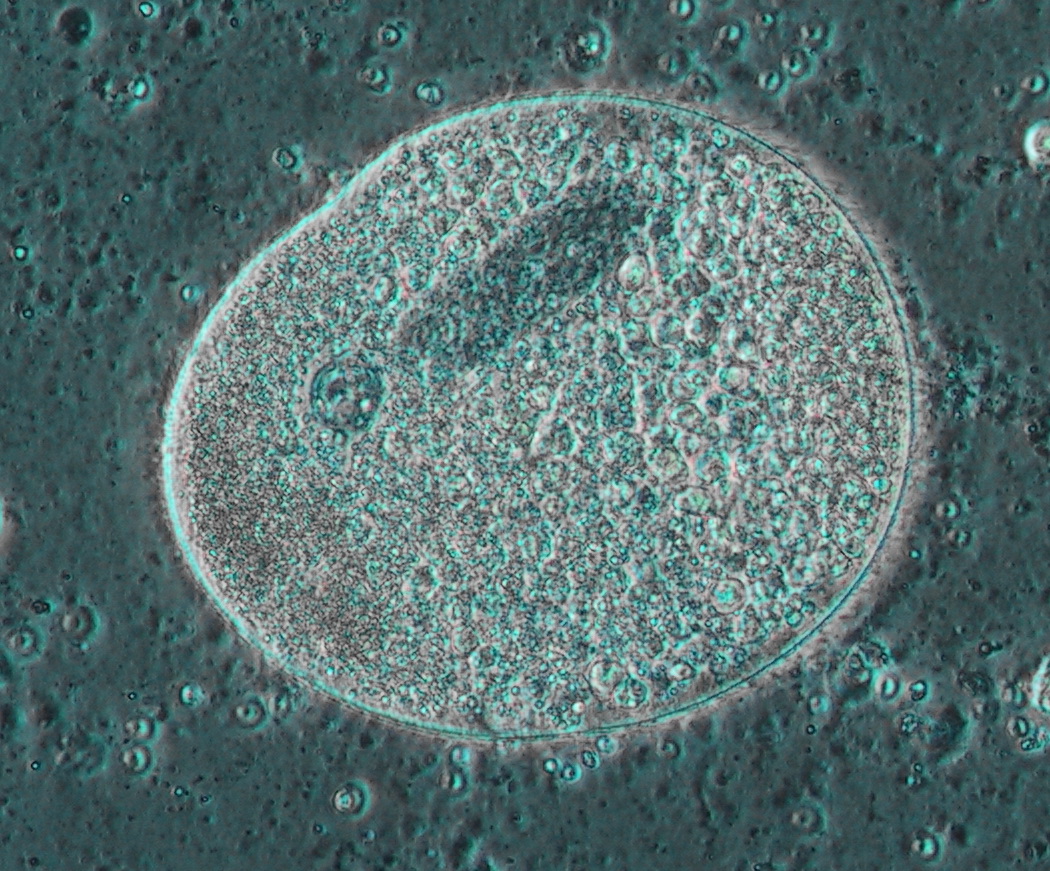
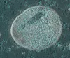 | database_122-2226_Ichthyo.jpg | All Pathogens, Fact Sheet Images | |
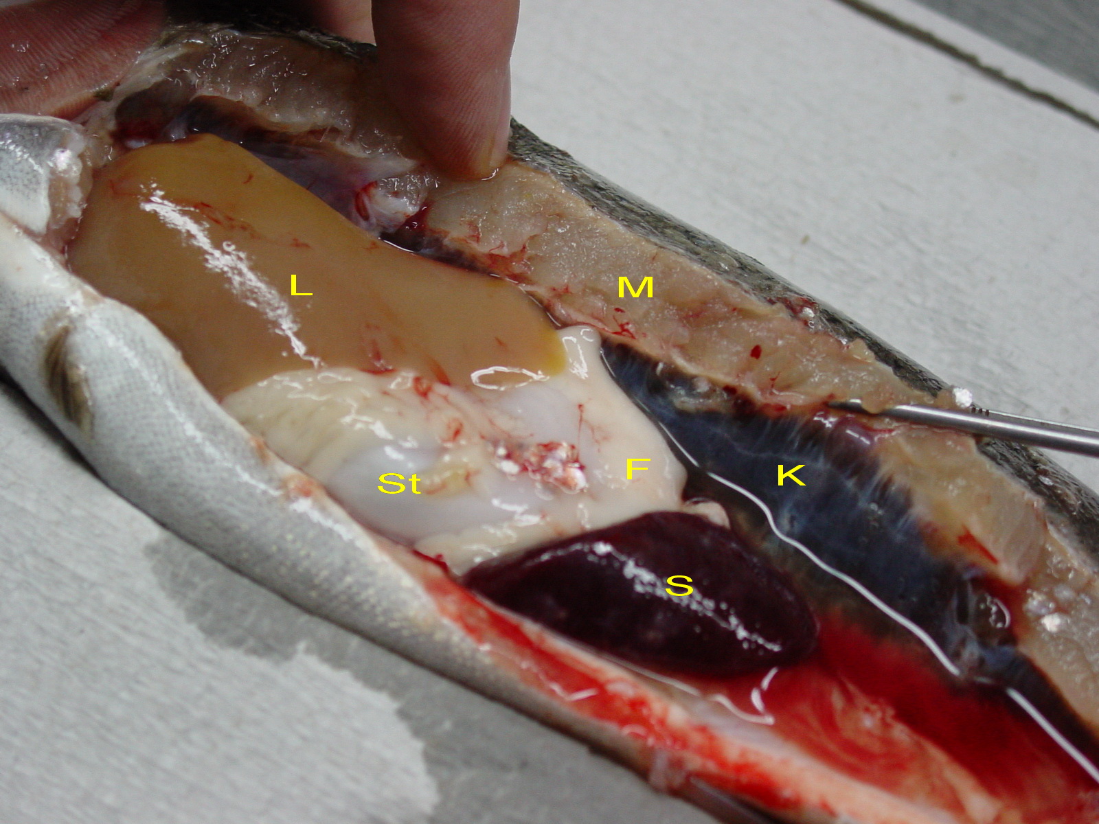
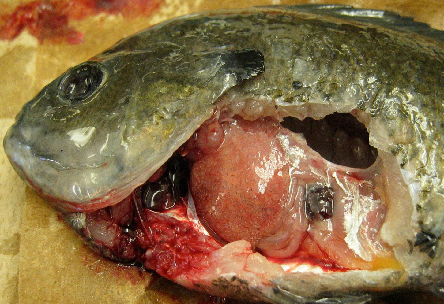
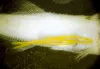
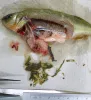
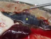 | database_DSC00517_pkd.jpg | All Pathogens, Fact Sheet Images | rainbow trout |
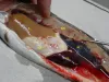 | database_DSC00518_pkd.jpg | All Pathogens, Fact Sheet Images | rainbow trout |
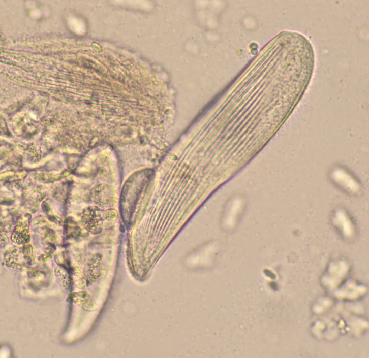
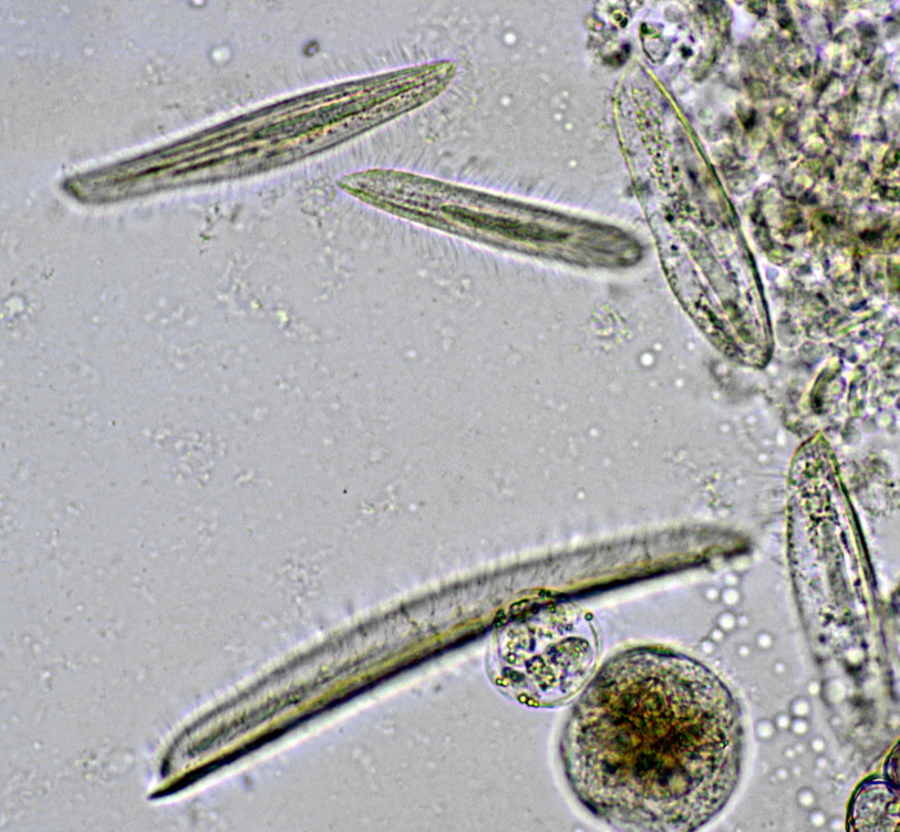
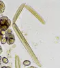
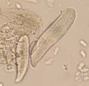 | database_SLH0027 - Free ciliates in seawater x20.jpg | All Pathogens, Fact Sheet Images | |
 | database_SLH0028 - Double infection.jpg | All Pathogens, Fact Sheet Images | |
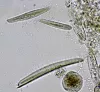 | database_SLH0046 - Ciliates double infection x20.jpg | All Pathogens, Fact Sheet Images | |
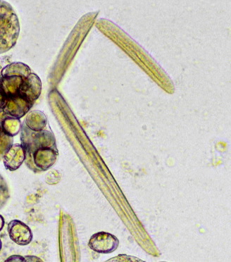
 | SLH0052 - linea.jpg | All Pathogens, Fact Sheet Images | |
 | FishPathogens_Cryptobia_IMG_8231.jpg | All Pathogens, Fact Sheet Images | |
 | BO_Dermocystidium_x63_25.jpg | All Pathogens, Fact Sheet Images | |
 | Renibacterium_2.png | All Pathogens, Fact Sheet Images | |
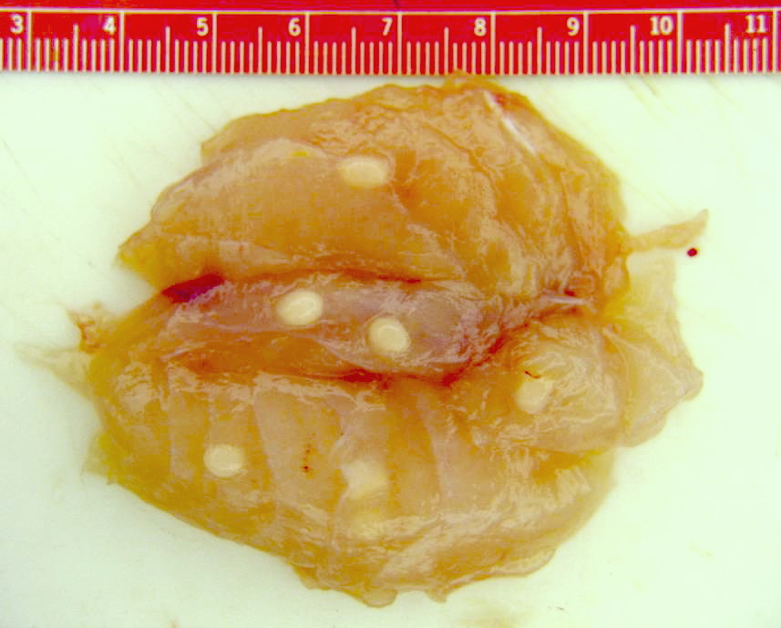
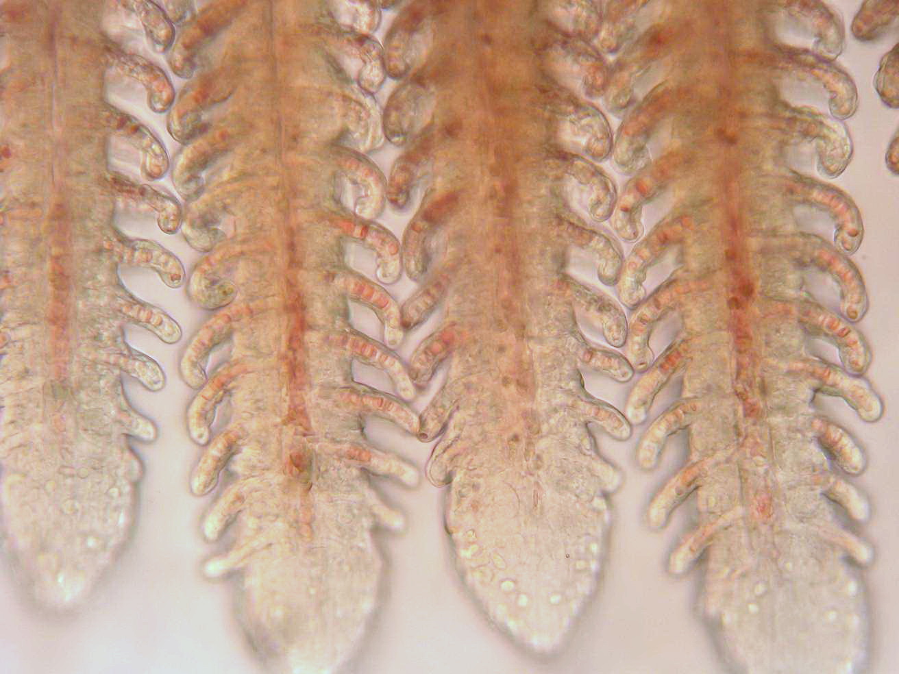
 | Clinostomum_03.jpg | All Pathogens, Top 10, Fact Sheet Images | |
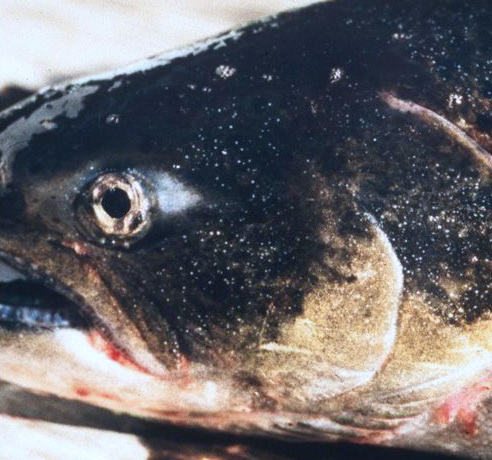
 | Henneguya_salminicola_cysts_02.jpg | All Pathogens, Fact Sheet Images, Top 10 | |
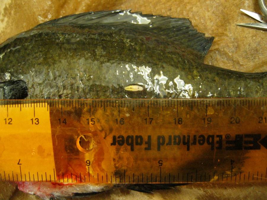
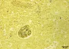
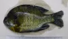
 | white_grub_01.jpg | All Pathogens, Fact Sheet Images, Top 10 | bluegill |
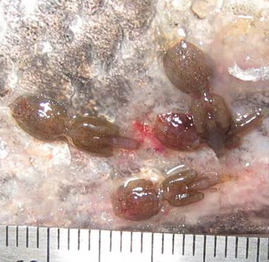
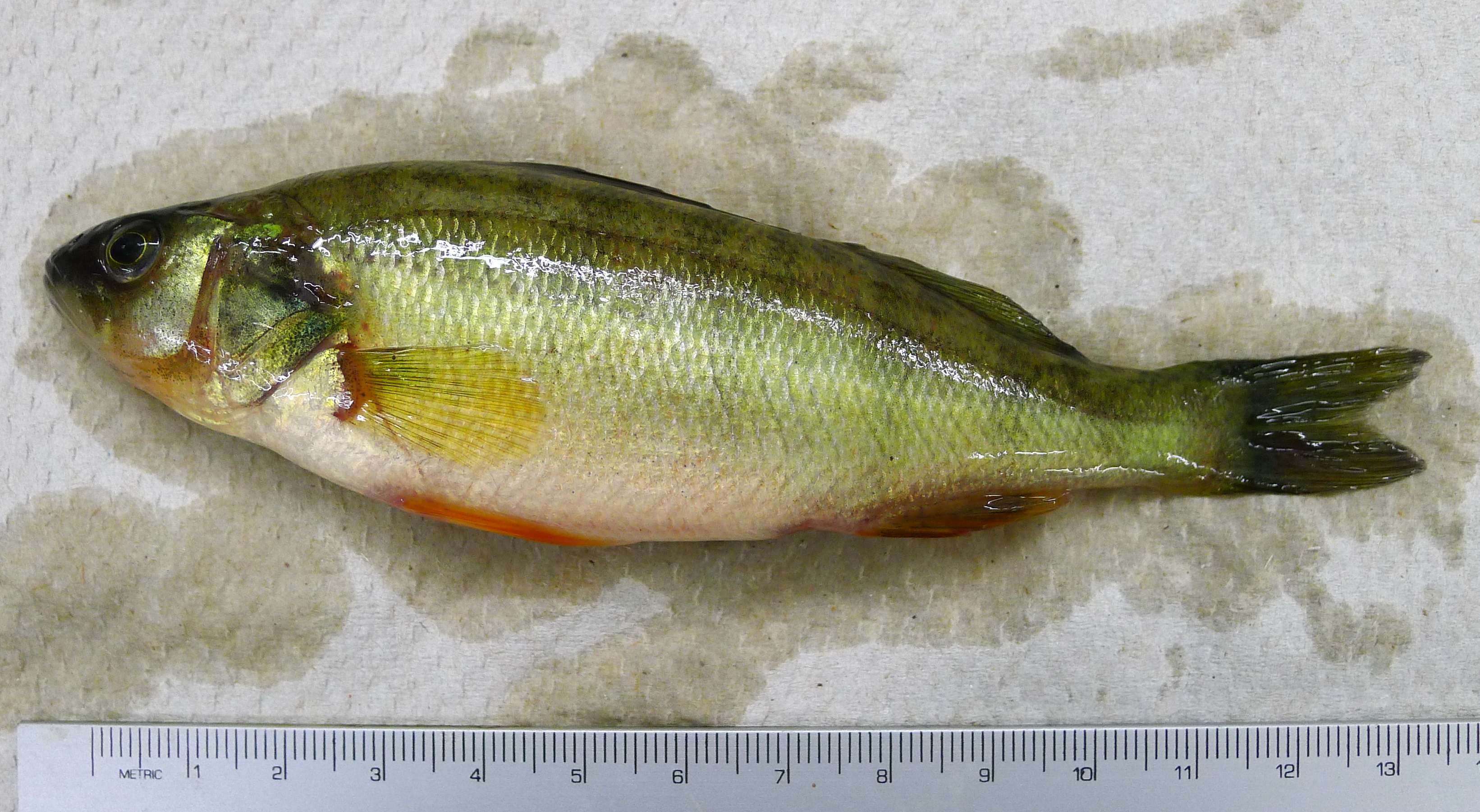
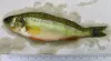
 | nanophyetus_salmonicola_CB09.jpg | All Pathogens, Top 10, Fact Sheet Images | yellow perch |
 | CB_IMG_0352.jpg | All Pathogens, Fact Sheet Images, Top 10, Hosts and other organisms | brown trout |
 | sealice_Lepiophthirius_02_crop.jpg | All Pathogens, Fact Sheet Images, Top 10 | |
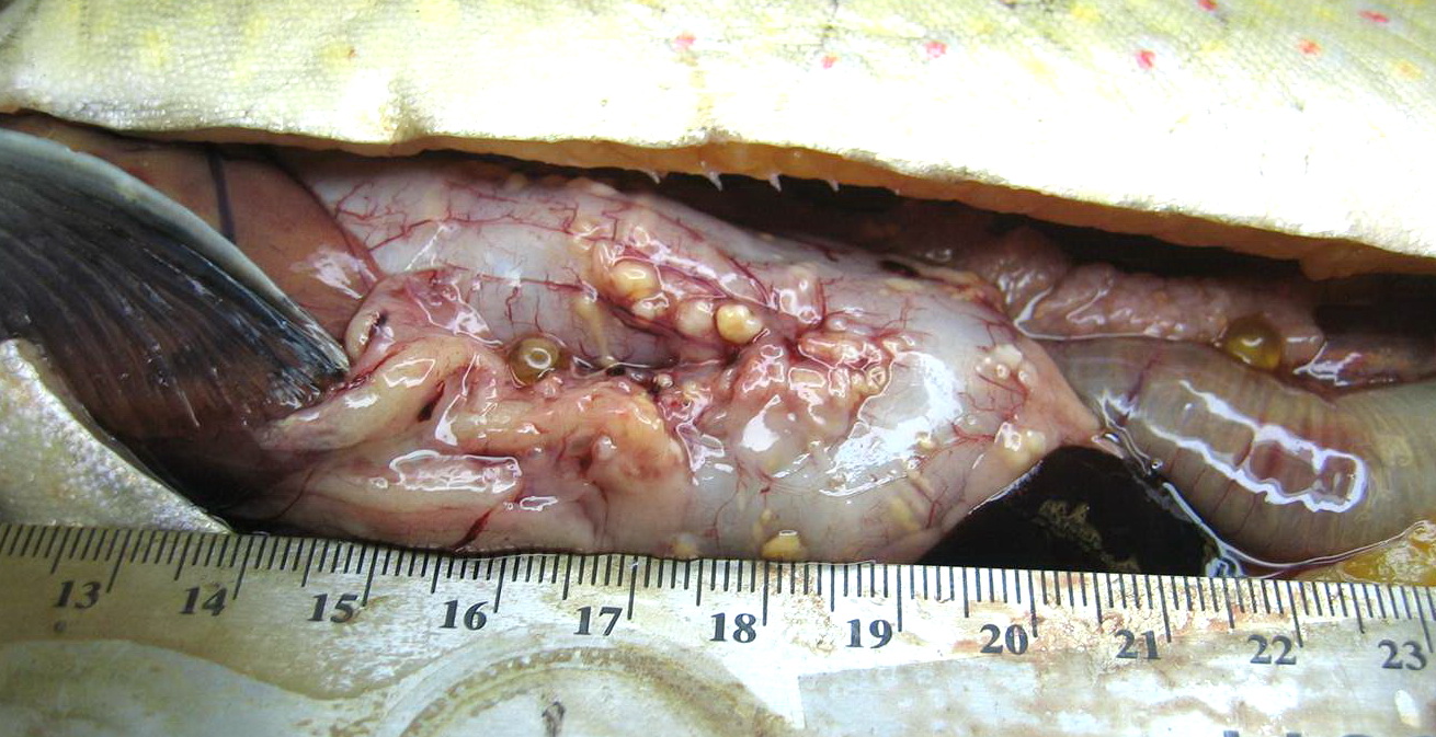
 | cesode_Diphyllobothrium_sp_CB03.jpg | All Pathogens, Fact Sheet Images, Top 10 | |
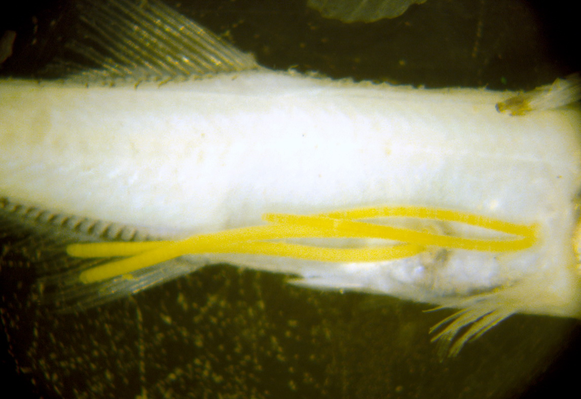
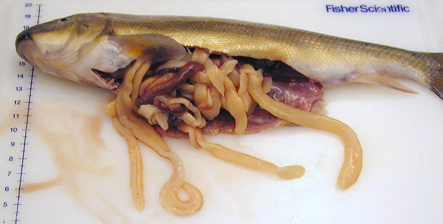
 | cestode_Lingula_intestinalis_CB01.jpg | All Pathogens, Fact Sheet Images, Top 10 | |
 | CB_adult_Ich_ChF.jpg | All Pathogens, Fact Sheet Images, Top 10 | Fall Chinook salmon |
 | SLH_DSC00179.jpg | All Pathogens, Top 10 | rainbow trout |
 | normal_gill.jpg | Fact Sheet Images, Hosts and other organisms | rainbow trout |
 | juga_sp.jpg | Fact Sheet Images, Hosts and other organisms | Juga snail |
| | CB_IMG_0352.jpg | All Pathogens, Fact Sheet Images, Top 10, Hosts and other organisms | brown trout |
 | P1000320.jpg | Fact Sheet Images, Hosts and other organisms | shrew (unspecified) |
 | P1000324.jpg | Fact Sheet Images, Hosts and other organisms | shrew (unspecified) |
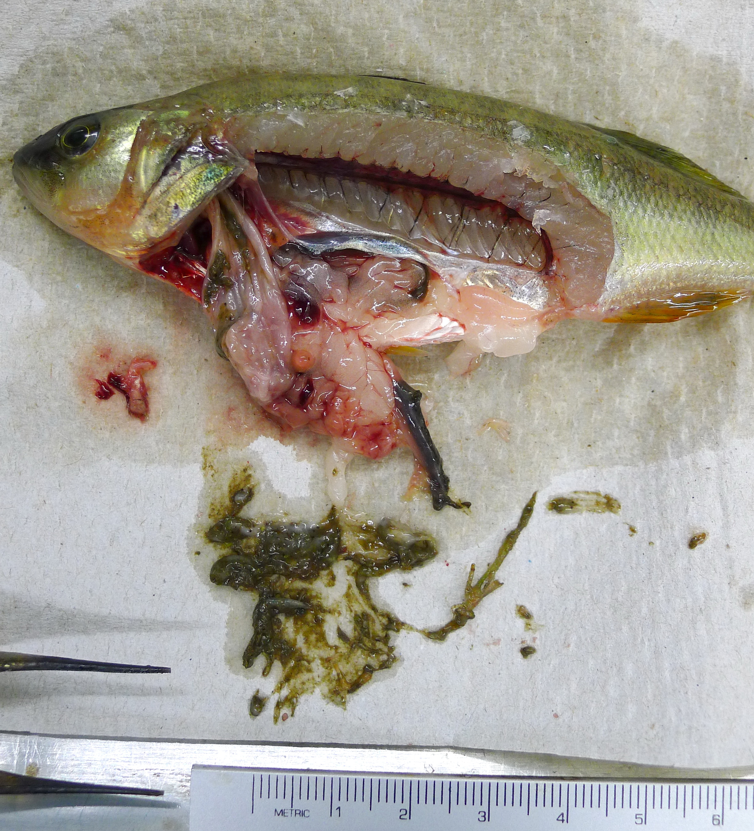
 | P1020299.jpg | Fact Sheet Images, Hosts and other organisms | yellow perch |
 | P1020300.jpg | Hosts and other organisms, Fact Sheet Images | yellow perch |
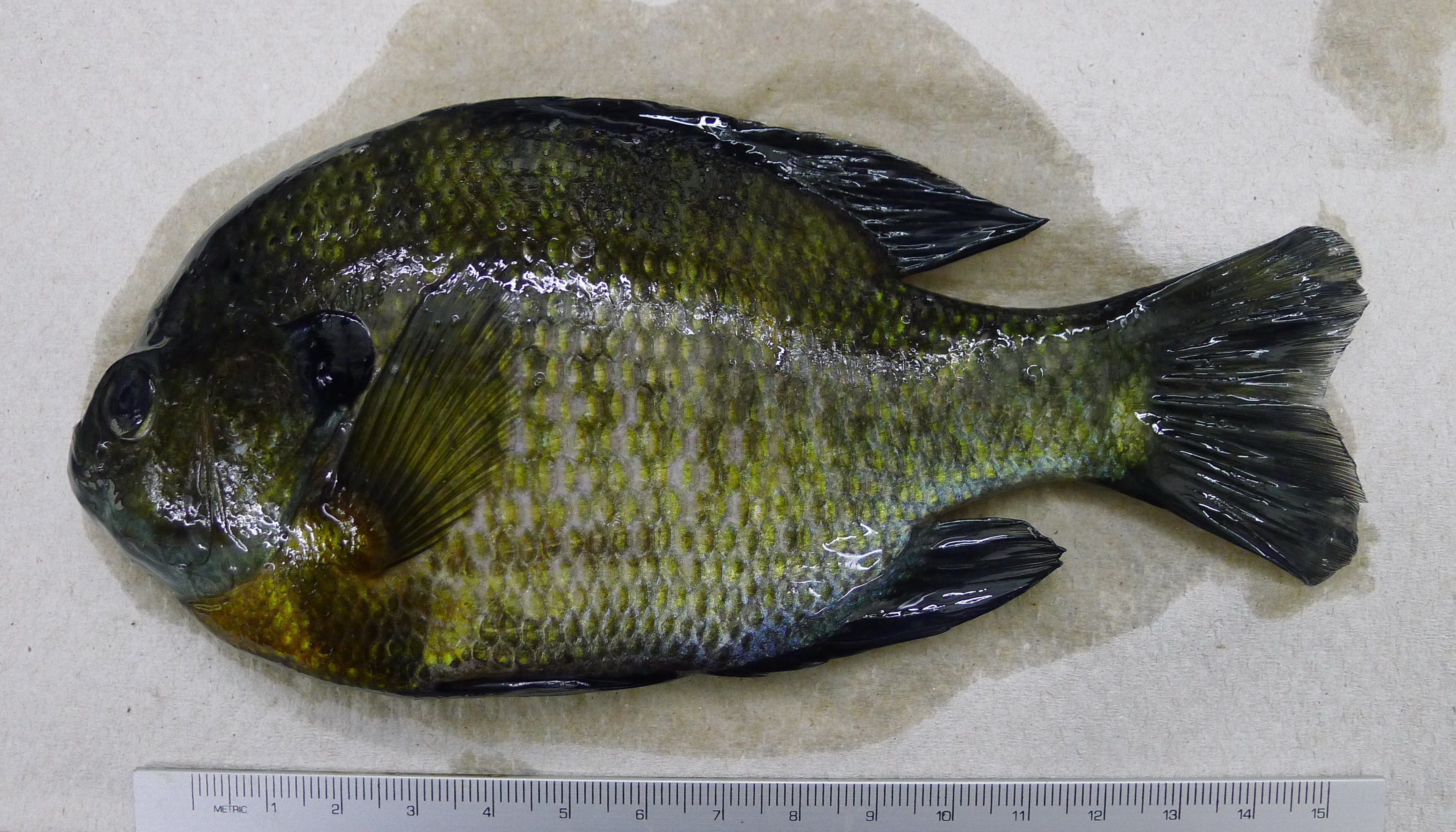
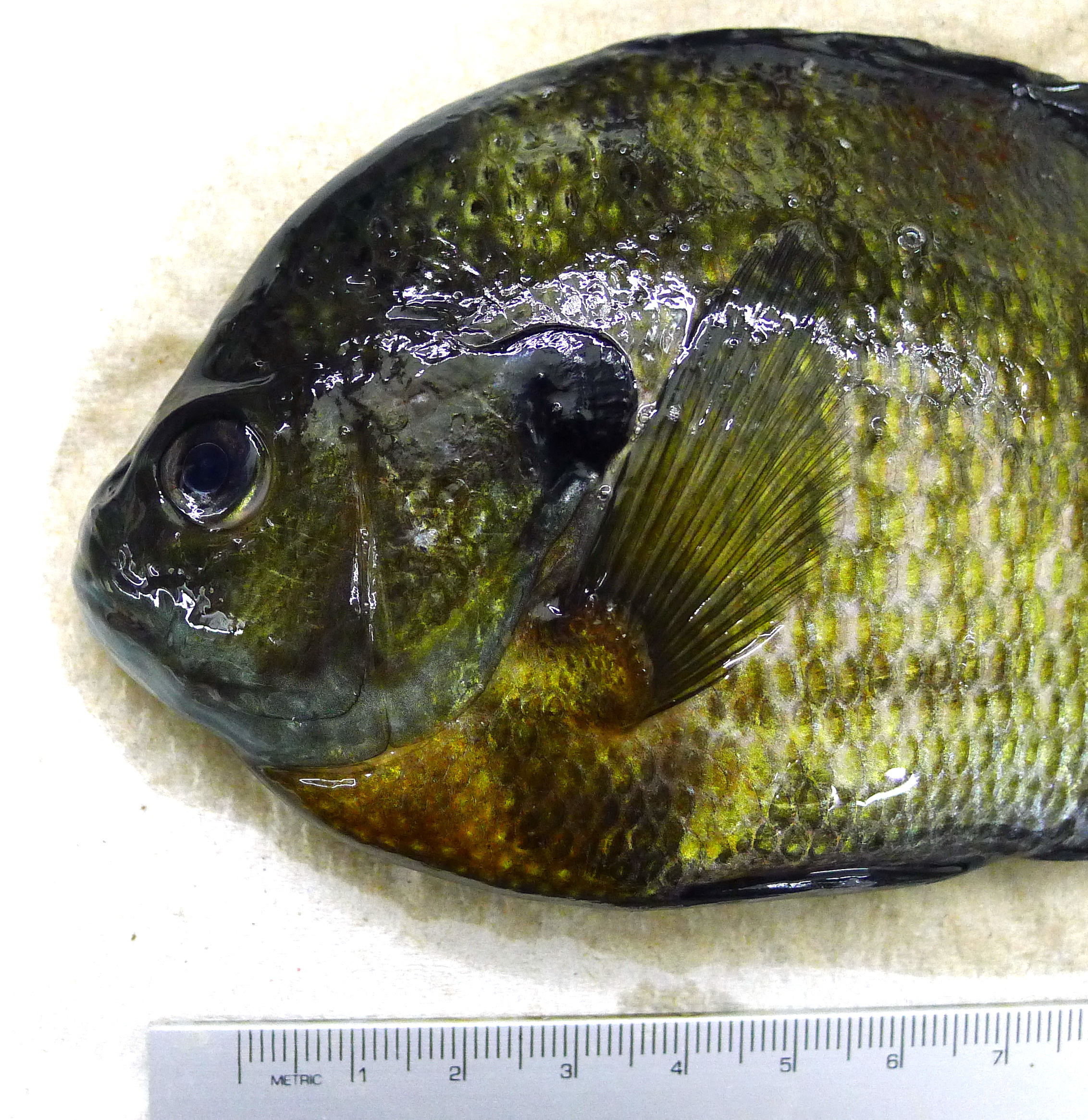
 | P1020301.jpg | Fact Sheet Images, Hosts and other organisms | bluegill |
 | P1020302.jpg | Fact Sheet Images, Hosts and other organisms | bluegill |