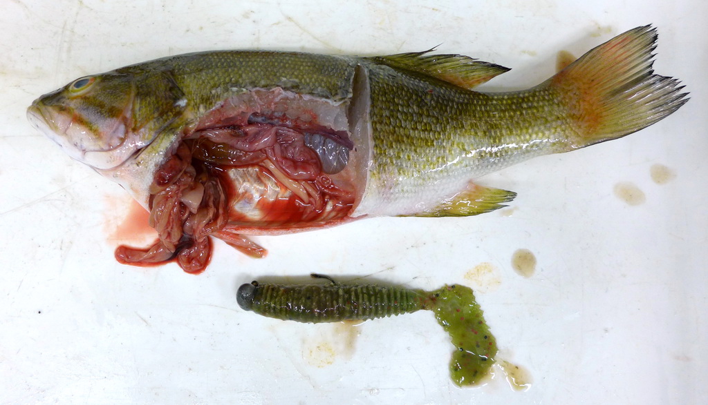
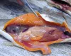
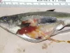
| P1000382ed.jpg | All Pathogens, Fact Sheet Images | smallmouth bass |
 | P1000380ed.jpg | All Pathogens, Fact Sheet Images | smallmouth bass |
 | P1000377ed.jpg | All Pathogens, Fact Sheet Images | smallmouth bass |
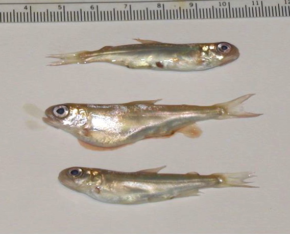
 | P1000375ed.jpg | All Pathogens, Fact Sheet Images | smallmouth bass |
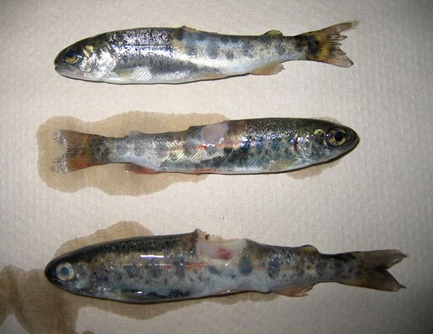
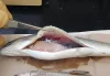
 | P1000368ed.jpg | All Pathogens, Hosts and other organisms, Fact Sheet Images | smallmouth bass |
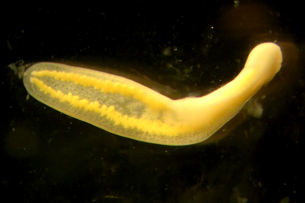
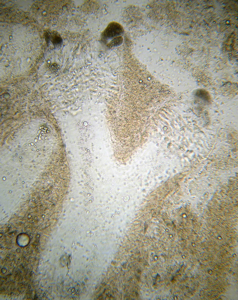
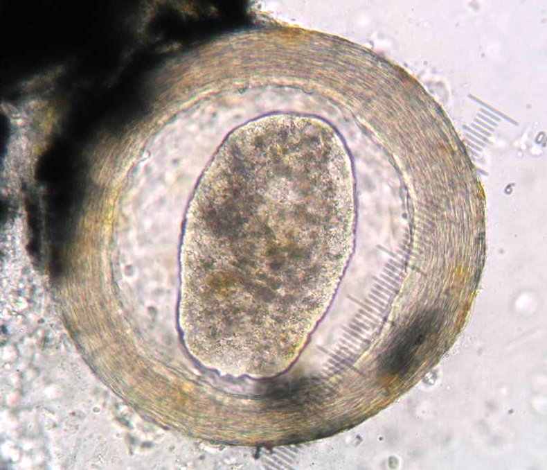
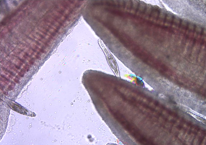
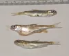
 | P1000365_db.jpg | All Pathogens, Fact Sheet Images | |
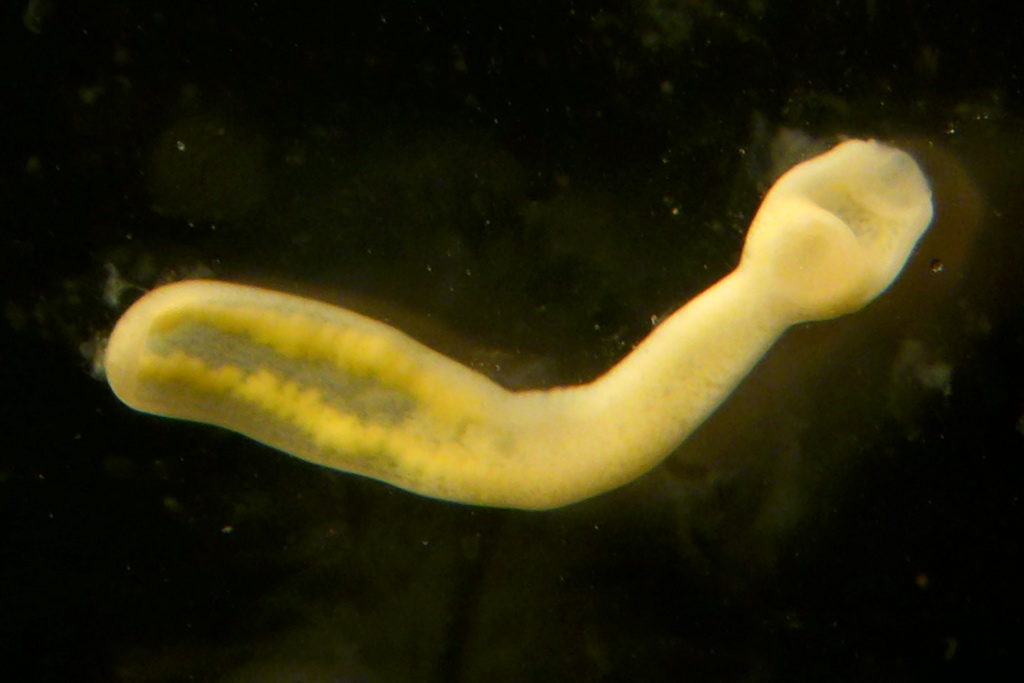
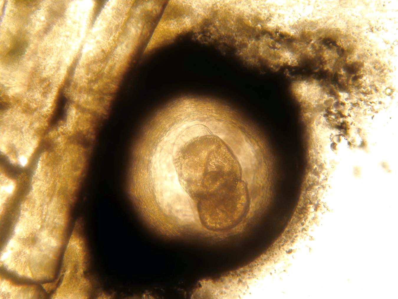
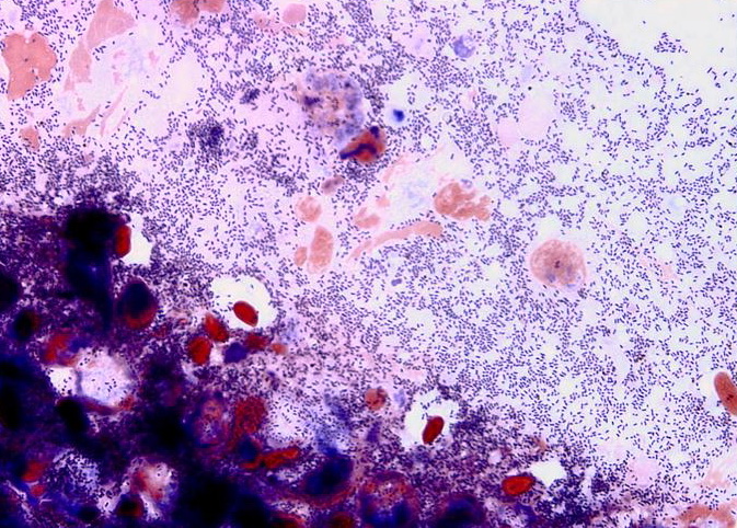
 | P1000366_db.jpg | All Pathogens, Fact Sheet Images | |
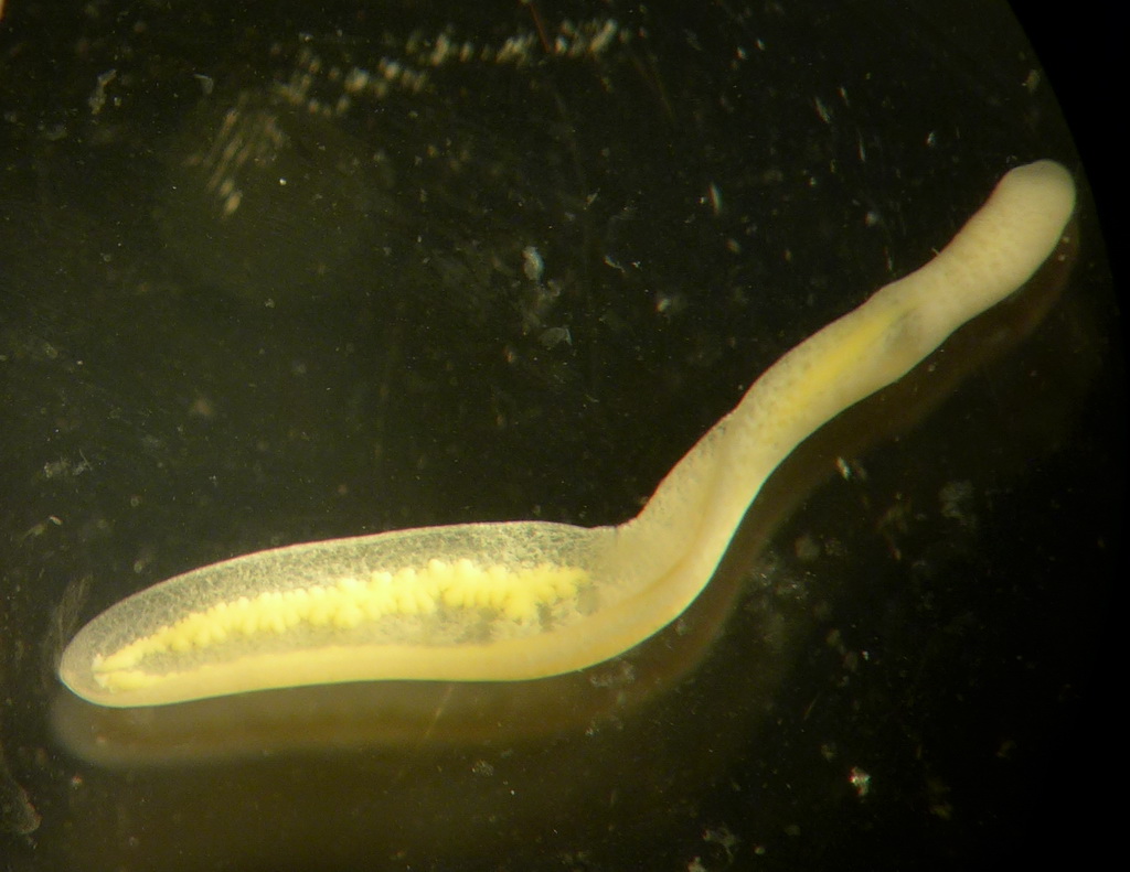
 | P1000367_db.jpg | All Pathogens, Fact Sheet Images | |
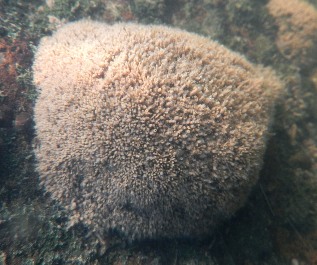
 | JCBoyle_june2012.jpg | Hosts and other organisms, Fact Sheet Images | Manayunkia sp. polychaete worm |
 | JCBoyle_june2012b.jpg | Hosts and other organisms, Fact Sheet Images | Manayunkia sp. polychaete worm |
 | TOH_june2012.jpg | Hosts and other organisms, Fact Sheet Images | Manayunkia sp. polychaete worm |
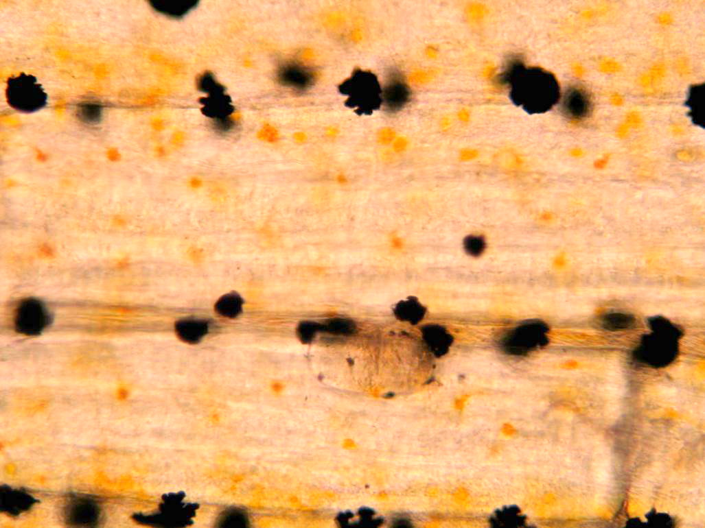
 | Neascus_02.jpg | All Pathogens, Fact Sheet Images | |
 | Neascus_05.jpg | All Pathogens, Fact Sheet Images | |
 | Neascus_06.jpg | All Pathogens, Fact Sheet Images | |
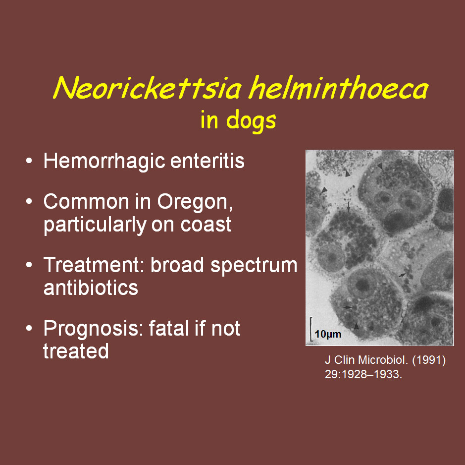
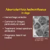 | CB_Neorickettsia_ppt.jpg | Fact Sheet Images, Illustration - organism | Nanophyetus |
 | CB_Philometroides_01.jpg | All Pathogens, Fact Sheet Images | largescale sucker |
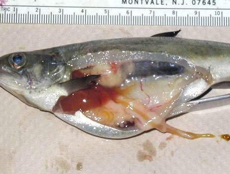
 | CB_Aeromonas_01.jpg | All Pathogens, Fact Sheet Images | rainbow trout |
 | CB_BKD_01.jpg | All Pathogens, Fact Sheet Images | |
 | CB_BKD_02_ChS.jpg | All Pathogens, Fact Sheet Images | Spring Chinook salmon |
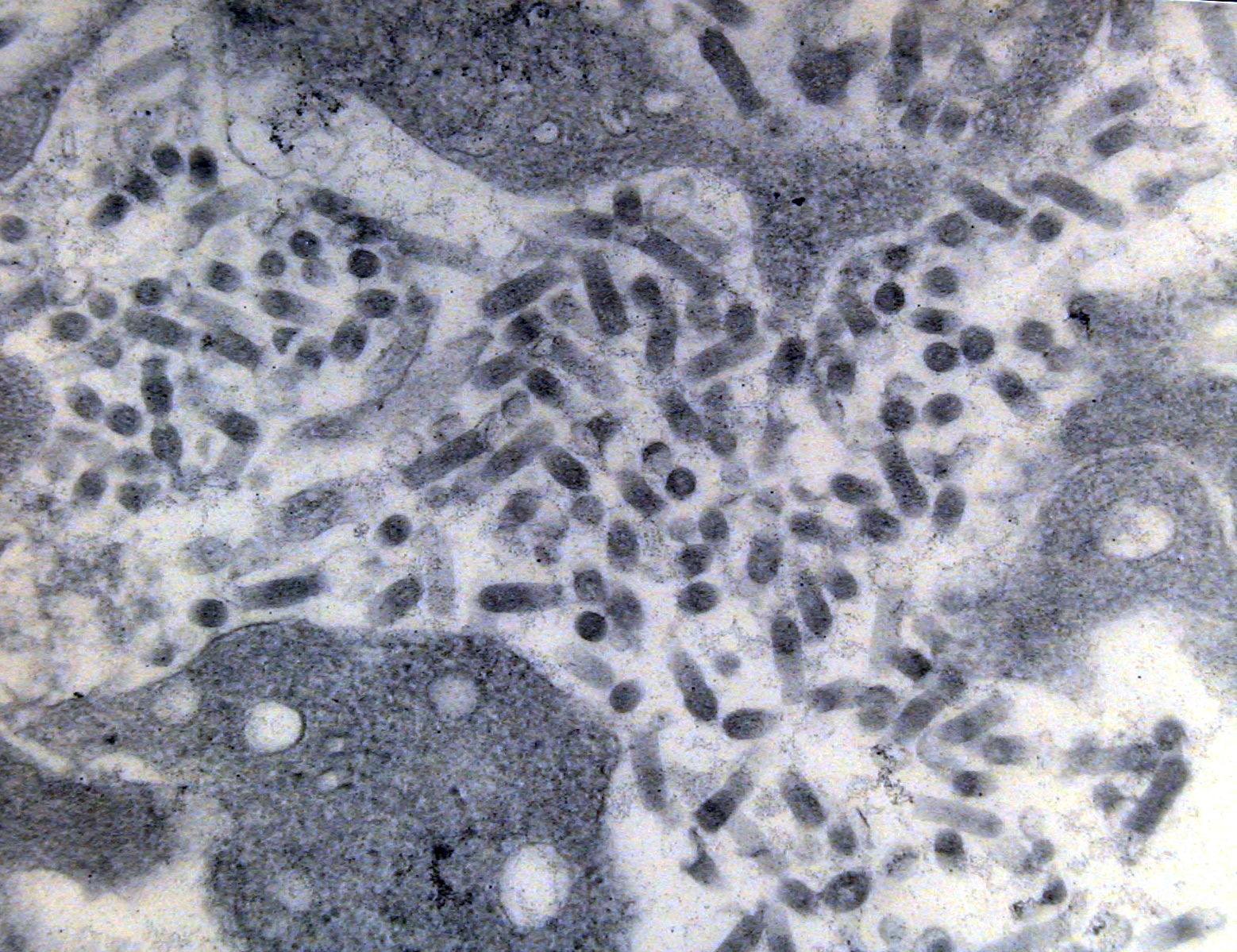
 | CB_BKD_03.jpg | All Pathogens, Fact Sheet Images | |
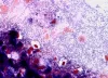 | CB_BKD_04.jpg | All Pathogens, Fact Sheet Images | |
 | CB_BKD_05.jpg | All Pathogens, Fact Sheet Images | |
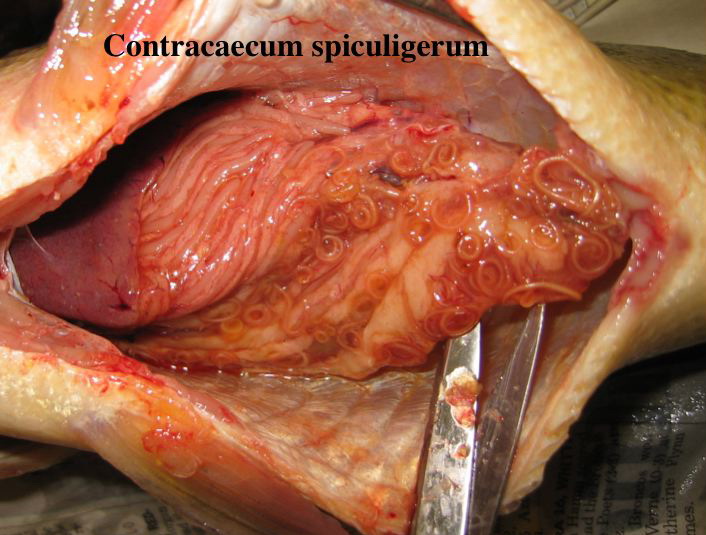
 | CB_Contracecum_01.jpg | All Pathogens, Fact Sheet Images | |
 | CB_CWD_01_StH.jpg | All Pathogens, Fact Sheet Images | steelhead |
 | CB_Dactylogyrus01.jpg | All Pathogens, Fact Sheet Images | |
 | VHSV_kaufman.jpg | All Pathogens, Fact Sheet Images | |
 | IHNV_kaufman1.jpg | All Pathogens, Fact Sheet Images | |
 | IHNV_kaufman2.jpg | All Pathogens, Fact Sheet Images | |
 | IHNVCPEplaque1.jpg | All Pathogens, Fact Sheet Images | |
 | IHNVCPE.jpg | All Pathogens, Fact Sheet Images | |