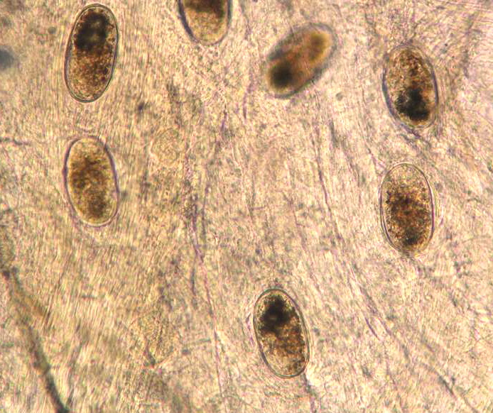
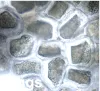
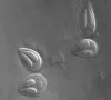
 | trematode_metacercaria_unidentified_CB03.jpg | All Pathogens, Fact Sheet Images | |
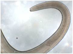
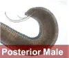
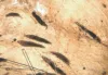
 | nematode_Anasakis_simplex_CB01.jpg | All Pathogens, Fact Sheet Images | |
 | nematode_Anasakis_simplex_CB02.jpg | All Pathogens, Fact Sheet Images | |
 | nematode_Hysterothylacium_sp_CB01.jpg | Illustration - organism, Fact Sheet Images | |
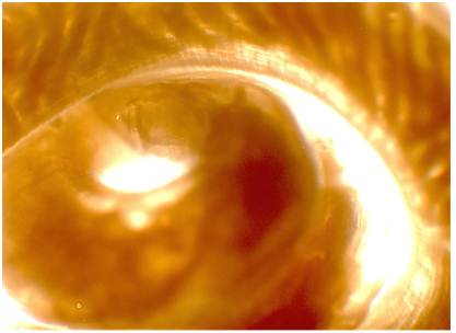
 | nematode_Hysterothylacium_sp_CB02.jpg | All Pathogens, Fact Sheet Images | |
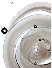
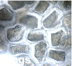
 | nematode_Hysterothylacium_sp_CB03.jpg.png | All Pathogens, Fact Sheet Images | |
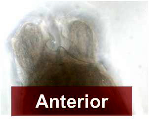
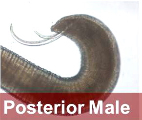
 | nematode_Hysterothylacium_sp_CB04.jpg | Illustration - organism, Fact Sheet Images | |
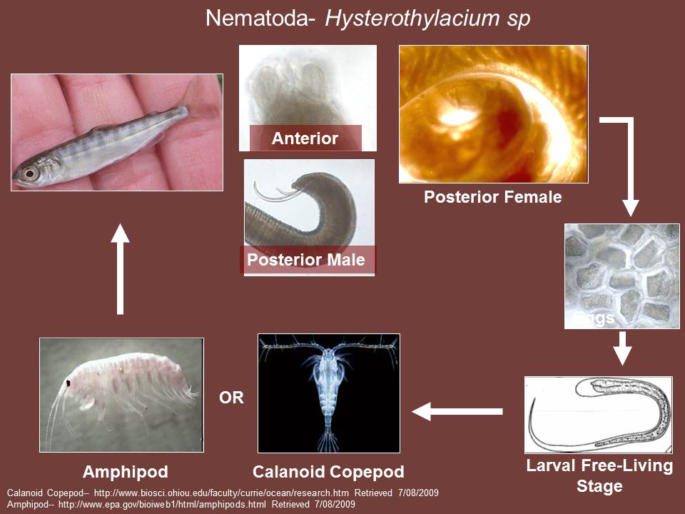
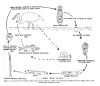
 | nematode_Hysterothylacium_sp_CB05.jpg | Illustration - life cycle, Fact Sheet Images | |
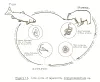
 | clinostomum_lifecycle.jpg | Illustration - life cycle, Fact Sheet Images | |
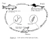
 | diphyllobothrium_lifecycle.gif | Illustration - life cycle, Fact Sheet Images | |
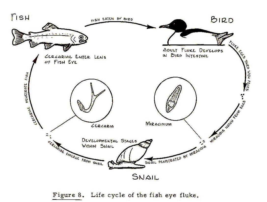
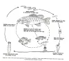
 | eyefluke_lifecycle.jpg | Illustration - life cycle, Fact Sheet Images | |
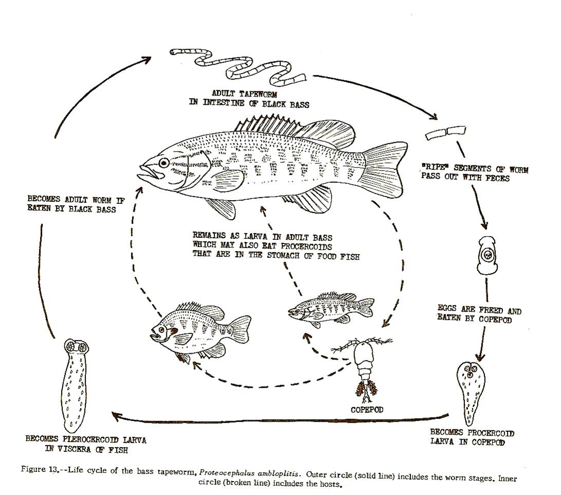
 | proteocephalus_lifecycle2.jpg | Illustration - life cycle, Fact Sheet Images | |
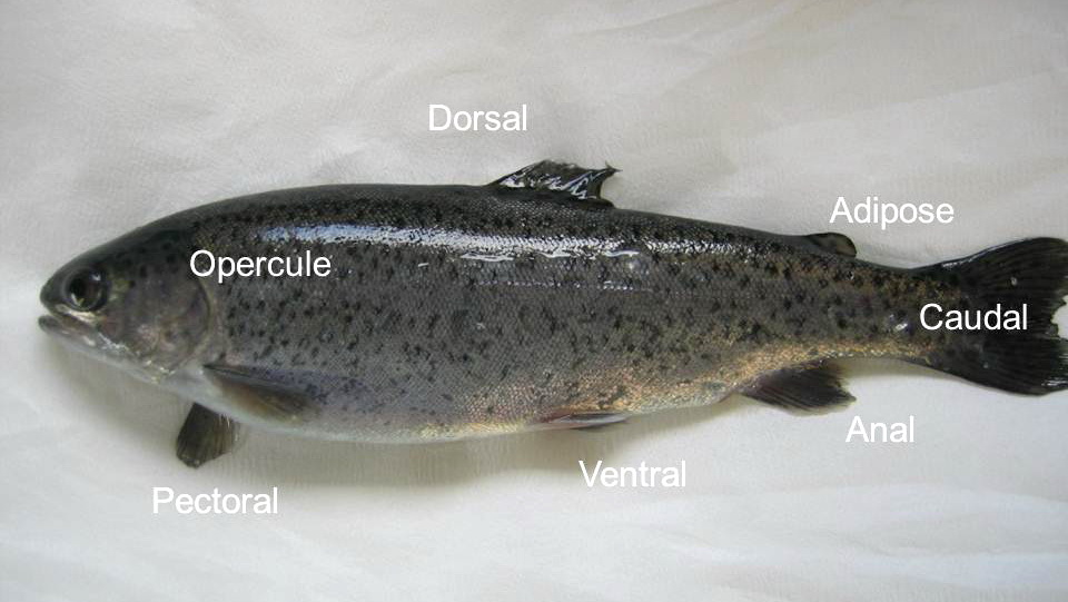
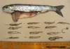
 | fish_fin_names_CB01.jpg | Hosts and other organisms, Fact Sheet Images | rainbow trout |
 | trichophyra_CB03.jpg | All Pathogens, Fact Sheet Images | |
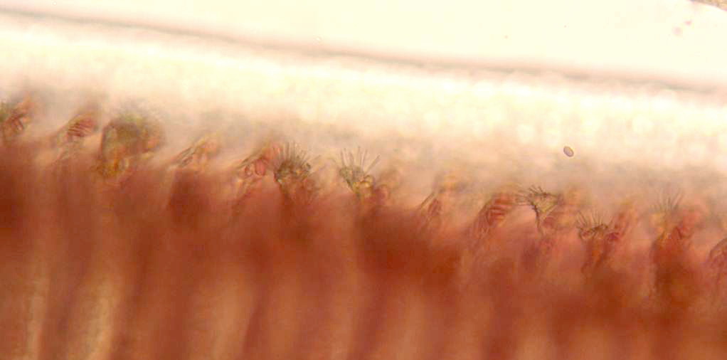
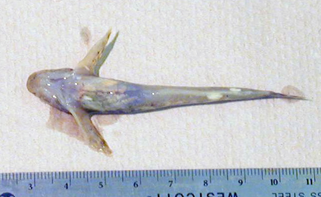
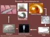
 | dermocystidium_CB01.jpg | All Pathogens, Fact Sheet Images | Fall Chinook salmon |
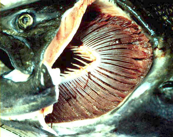
 | dermocystidium_CB02.jpg | All Pathogens, Fact Sheet Images | Fall Chinook salmon |
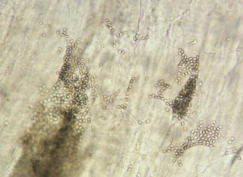
 | dermocystidium_CB03.jpg | All Pathogens, Fact Sheet Images | Fall Chinook salmon |
 | nitzschia_CB01.jpg | All Pathogens, Fact Sheet Images | white sturgeon |
 | Minsidiosus_CB02.jpg | All Pathogens, Fact Sheet Images | |
 | Minsidiosus_CB01.jpg | All Pathogens, Fact Sheet Images | |
 | Minsidiosus_CB03.jpg | All Pathogens, Fact Sheet Images | |
 | glugea_CB01.jpg | All Pathogens, Fact Sheet Images | sculpin (unspecified) |
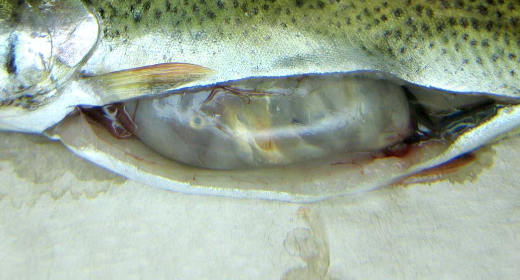
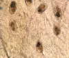
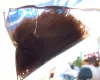
 | stomach_content_CB01.jpg | Fact Sheet Images, Hosts and other organisms | brown trout |
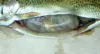 | stomach_content_CB02.jpg | Fact Sheet Images, Hosts and other organisms | brown trout |
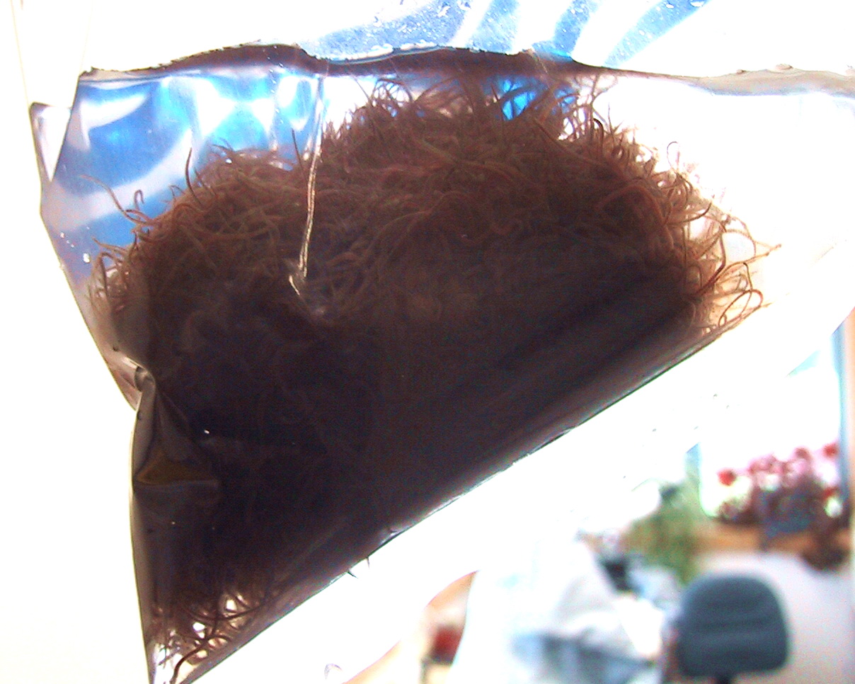
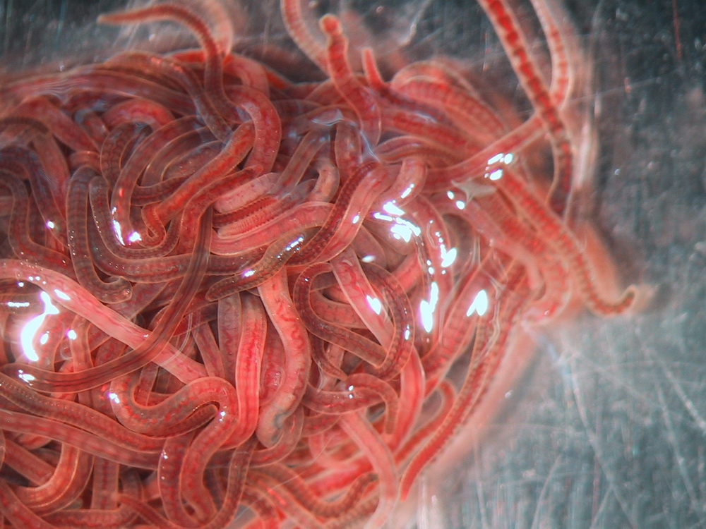
 | 171_7112_lumbriculus.jpg | Fact Sheet Images, Hosts and other organisms | Lumbriculus / black worms |
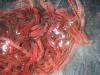 | 171_7113_lumbriculus.jpg | Fact Sheet Images, Hosts and other organisms | Lumbriculus / black worms |
 | DSC00263_amandi.jpg | Personel, Fact Sheet Images | |
 | DSC00494_bjork.jpg | Personel, Fact Sheet Images | |
 | DSC00495_arsan.jpg | Personel, Fact Sheet Images | |
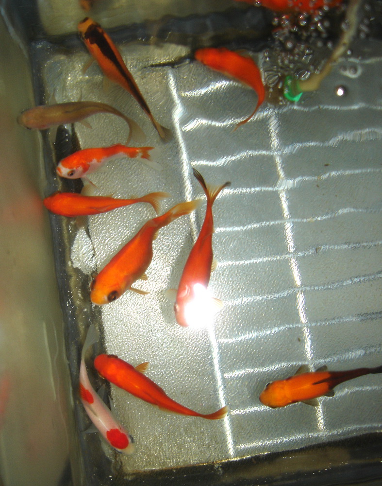
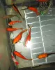 | DSC00538_goldfish.jpg | Fact Sheet Images, Hosts and other organisms | goldfish |