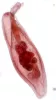
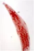
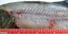
 | Acanthocephala_Echinorhynchus_lageniformis_CB01.jpg | All Pathogens, Fact Sheet Images | |
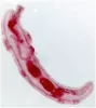
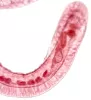
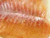
 | Acanthocephala_Echinorhynchus_lageniformis_CB04.jpg | All Pathogens, Fact Sheet Images | |
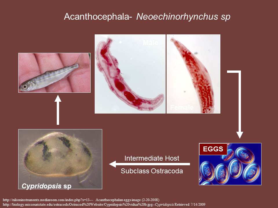
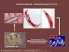 | Acanthocephala_Neoechinorhynchus_sp_CB03.jpg | Illustration - life cycle, Fact Sheet Images | |
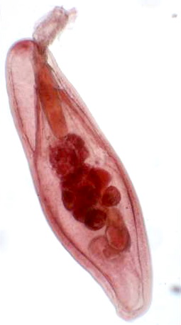
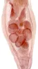
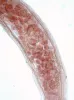
 | Acanthocephala_Neoechinorhynchus_sp_CB01.jpg | All Pathogens, Fact Sheet Images | |
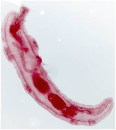
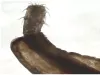
 | Acanthocephala_Neoechinorhynchus_sp_CB02.jpg | All Pathogens, Fact Sheet Images | |
 | Acanthocephala_Echinorhynchus_gadi_CB03.jpg | All Pathogens, Fact Sheet Images | |
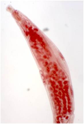
 | Acanthocephala_Neoechinorhynchus_sp_CB04.jpg | All Pathogens, Fact Sheet Images | |
 | Acanthocephala_Neoechinorhynchus_sp_CB05.jpg | All Pathogens, Fact Sheet Images | |
 | Acanthocephala_Neoechinorhynchus_sp_CB06.jpg | All Pathogens, Fact Sheet Images | |
 | Acanthocephala_Neoechinorhynchus_sp_CB07.jpg | All Pathogens, Fact Sheet Images | |
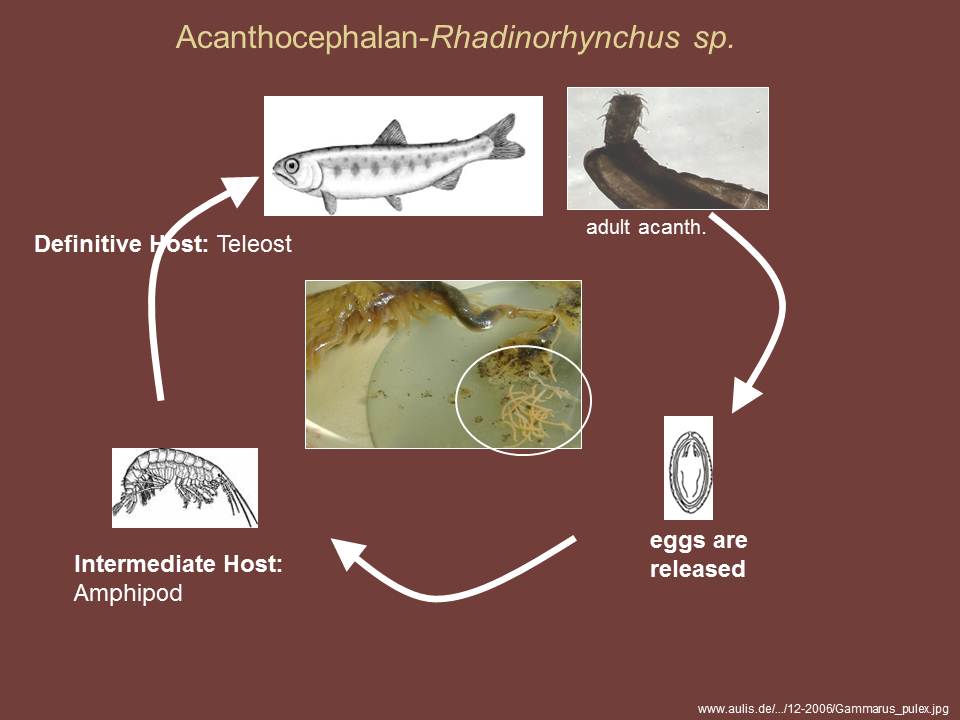
 | Acanthocephala_Rhadinorhynchus_sp_CB03.jpg | Illustration - life cycle, Fact Sheet Images | |
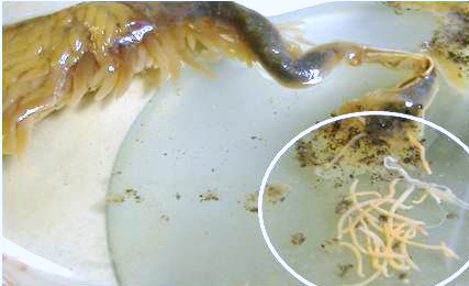
 | Acanthocephala_Rhadinorhynchus_sp_CB02.png | All Pathogens, Fact Sheet Images | |
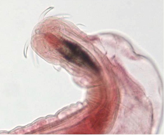
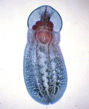
 | Acanthocephala_Rhadinorhynchus_sp_CB01.jpg | All Pathogens, Fact Sheet Images | |
 | trematode_CB01.jpg | All Pathogens, Fact Sheet Images | |
 | trematode_Posthodiplostomum_minimum_CB01.jpg | All Pathogens, Fact Sheet Images | |
 | trematode_Posthodiplostomum_minimum_CB03.jpg | All Pathogens, Fact Sheet Images | |
 | trematode_Clinostomum_marginatum_CB03.jpg | All Pathogens, Fact Sheet Images | |
 | trematode_Clinostomum_marginatum_CB04.jpg | All Pathogens, Fact Sheet Images | |
 | trematode_Clinostomum_marginatum_CB05.jpg | All Pathogens, Fact Sheet Images | |
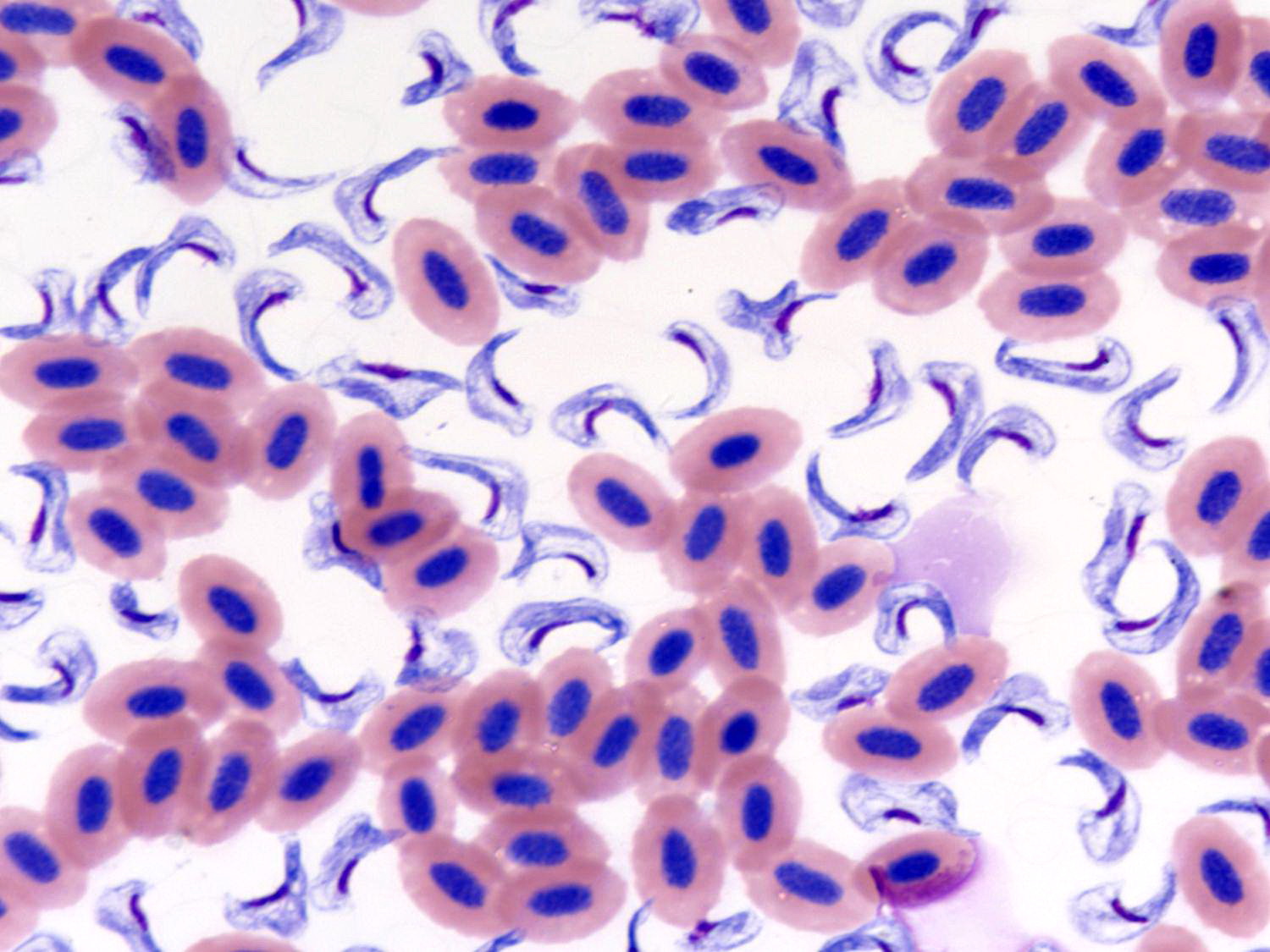
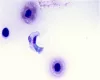 | hemoflagellate_Cryptobia_salmositica_CB01.jpg | All Pathogens, Fact Sheet Images | |
 | hemoflagellate_Cryptobia_salmositica_CB02.jpg | All Pathogens, Fact Sheet Images | |
| | hemoflagellate_Cryptobia_salmositica_CB02.jpg | All Pathogens, Fact Sheet Images | |
| | hemoflagellate_Cryptobia_salmositica_CB01.jpg | All Pathogens, Fact Sheet Images | |
 | hemoflagellate_Cryptobia_salmositica_CB03.jpg | All Pathogens, Fact Sheet Images | |
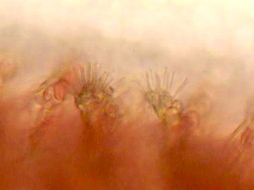
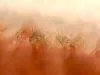 | tricophyra_CB01.jpg | All Pathogens, Fact Sheet Images | |
 | tricophyra_CB02.jpg | All Pathogens, Fact Sheet Images | |
 | trichodinella_CB01.jpg | All Pathogens, Fact Sheet Images | |
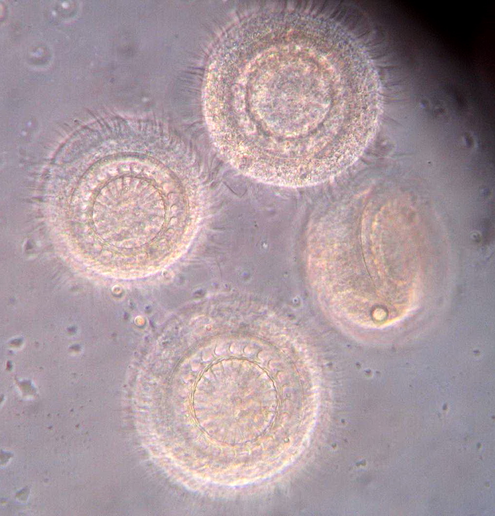
 | trichodina_CB01.jpg | All Pathogens, Fact Sheet Images | |
 | trichodina_CB02.jpg | All Pathogens, Fact Sheet Images | |
 | trichodina_CB03.jpg | All Pathogens, Fact Sheet Images | |