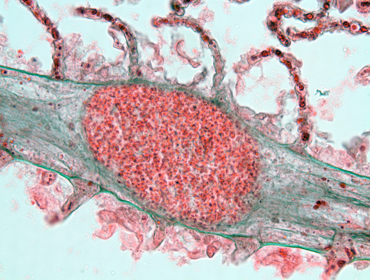
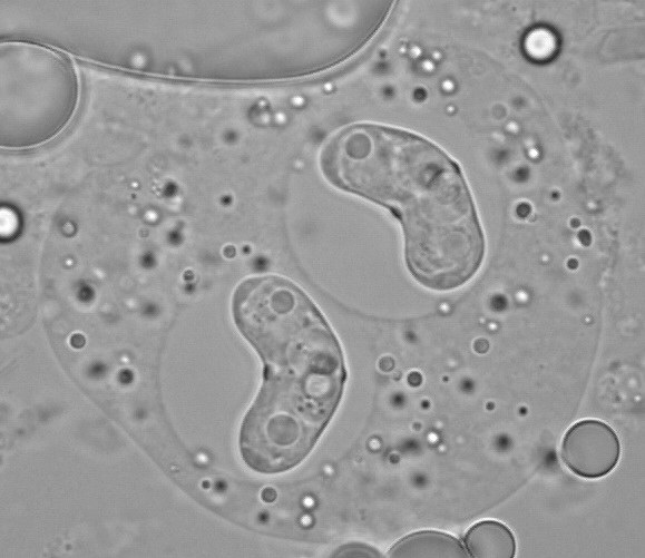
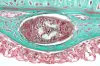
 | BO_Loma_x63_20.jpg | All Pathogens | |
 | CB_Kidtremyellow.jpg | All Pathogens, Fact Sheet Images | bluegill |
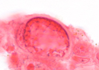
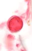
 | sanguinicola_CB04.jpg | All Pathogens, Fact Sheet Images | |
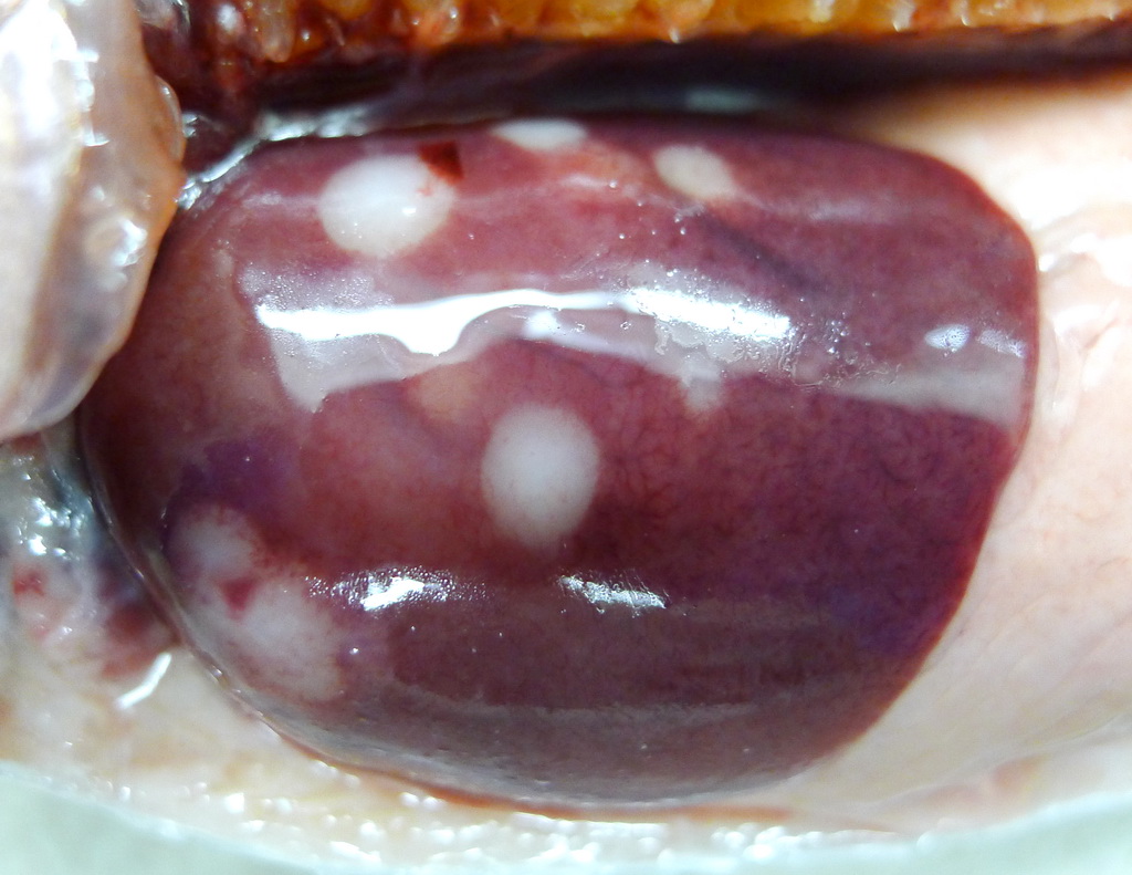
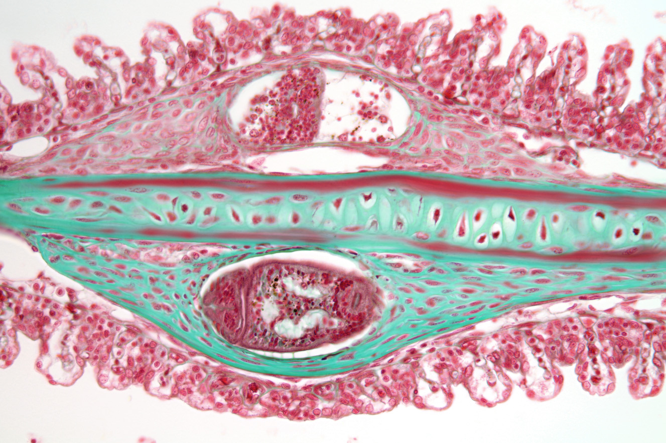
 | P1080678_Cshasta_liver.jpg | All Pathogens | rainbow trout |
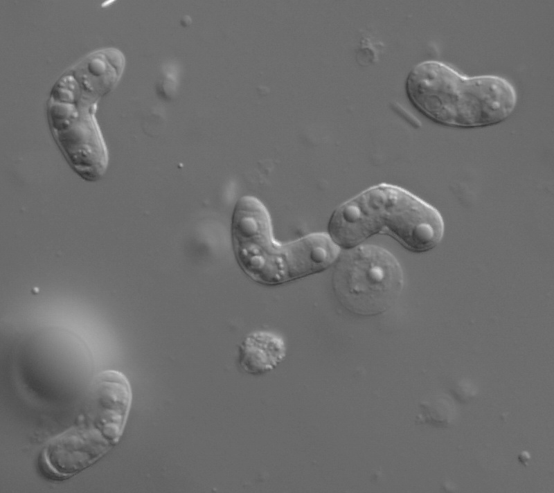
 | CB_IMG_0352.jpg | All Pathogens, Fact Sheet Images, Top 10, Hosts and other organisms | brown trout |
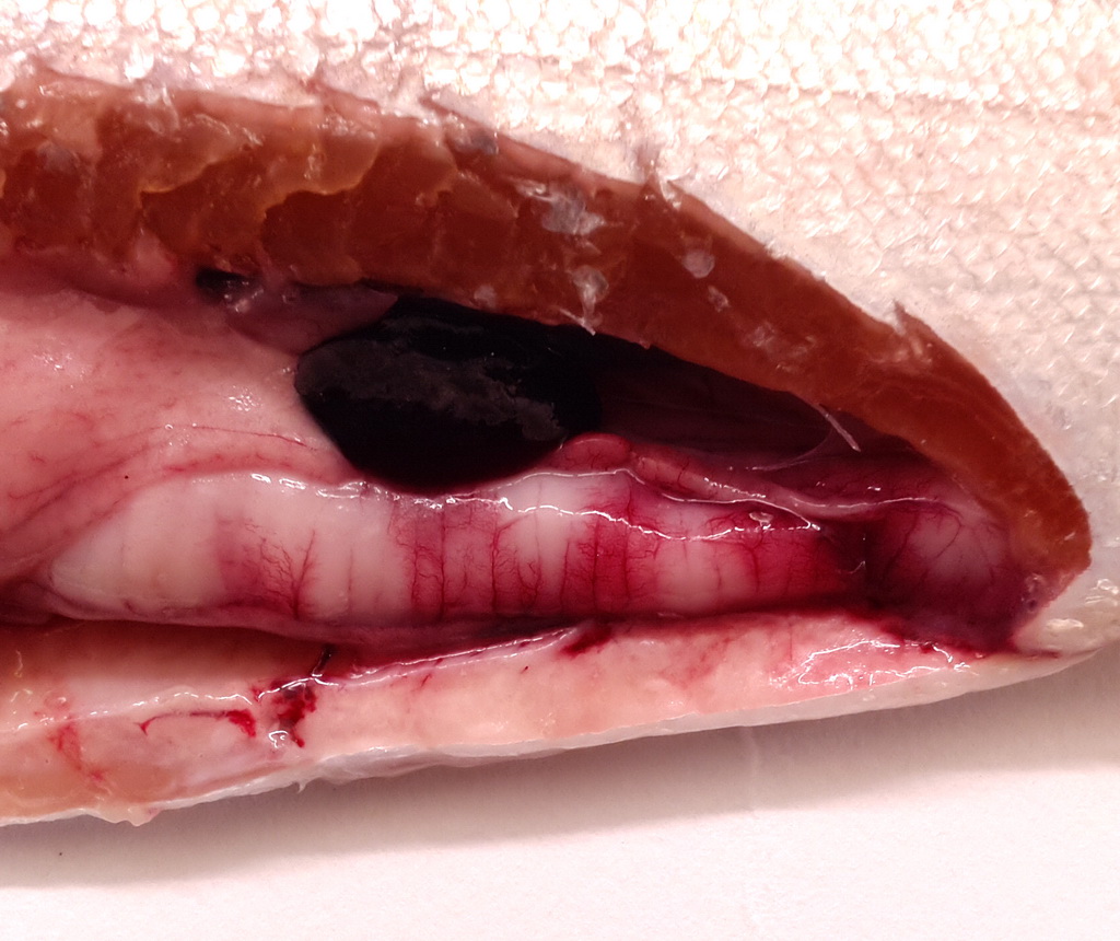
 | IMG_20130402_rbtCs2.jpg | All Pathogens | rainbow trout |
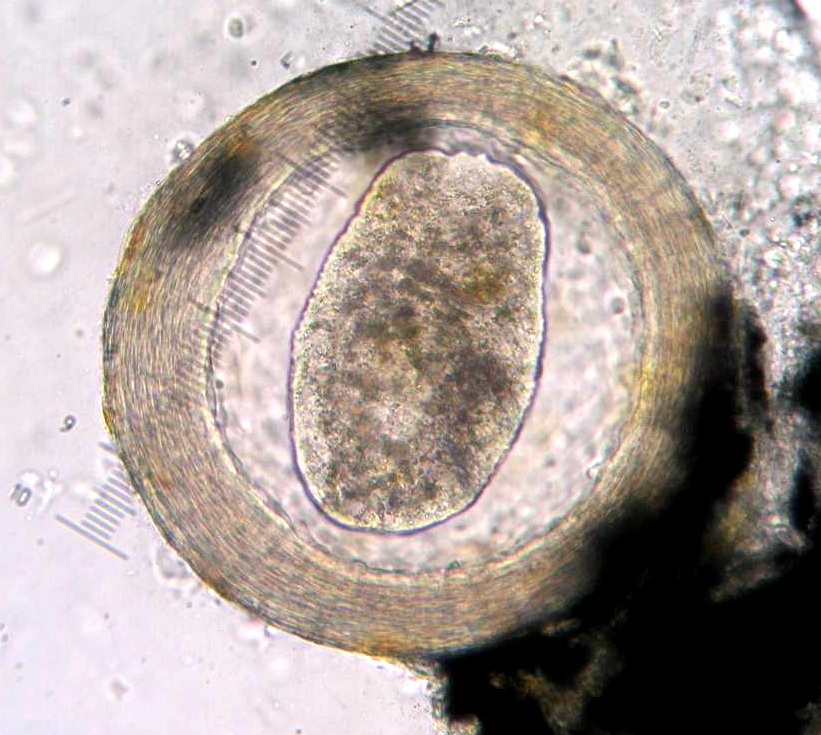
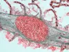
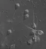
 | BO_miracidium_x63_12.jpg | All Pathogens | |
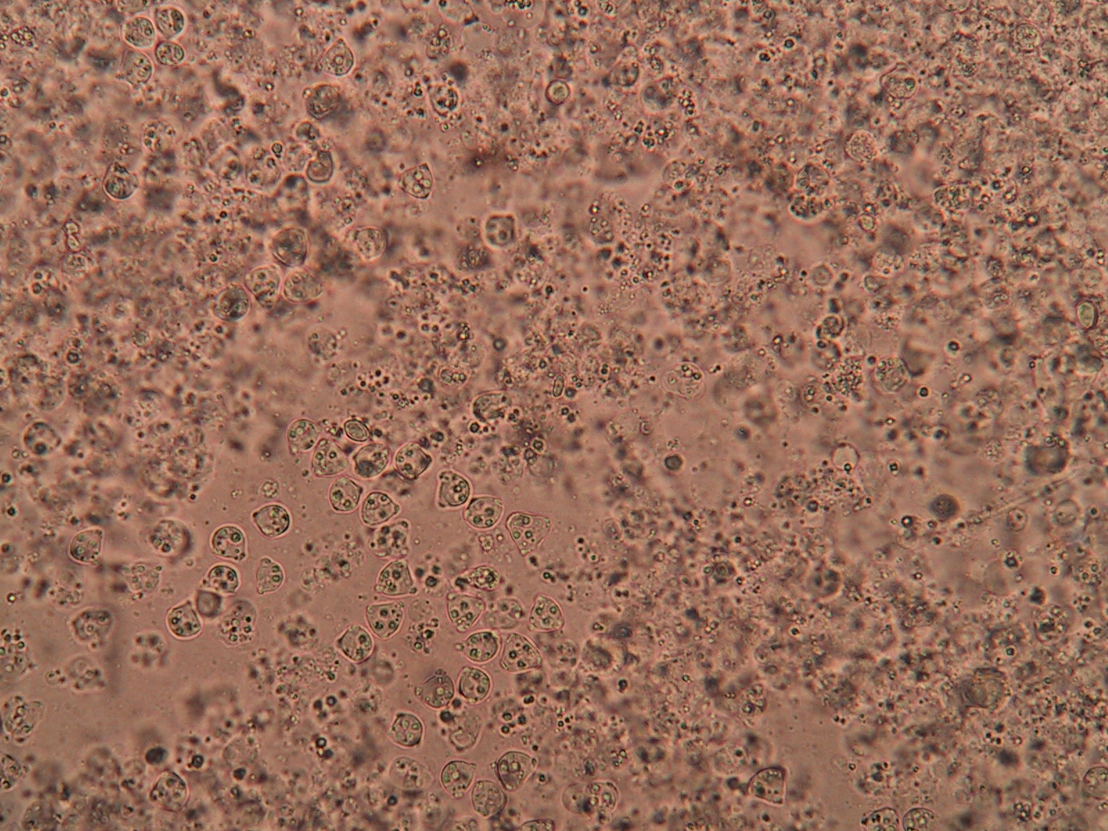
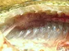
 | 177_7781.JPG | All Pathogens, Fact Sheet Images | |
 | sanguinicola_CB06.jpg | All Pathogens, Fact Sheet Images | |
 | BO_miracidium_x63_13.jpg | All Pathogens | |
 | sanguinicola_CB07.jpg | All Pathogens, Fact Sheet Images | |
 | Cshasta03.jpg | All Pathogens, Fact Sheet Images | |
 | sanguinicola_CB08.jpg | All Pathogens, Fact Sheet Images | |
 | OMV-Masu.jpg | All Pathogens, Fact Sheet Images | |
 | BO_trem_StH_x20_05.jpg | All Pathogens | steelhead |
 | sanguinicola_CB09.jpg | All Pathogens, Fact Sheet Images | |
 | L14m7709_1800b_100bf_1_3a.jpg | All Pathogens, Fact Sheet Images | |
 | BO_trem_StH_x20_06.jpg | All Pathogens | steelhead |
 | sanguinicola_CB10.jpg | All Pathogens, Fact Sheet Images | |
 | L14m7809_0900a_100nm_1_12a.jpg | All Pathogens, Fact Sheet Images | |
 | sanguinicola_CB11.jpg | All Pathogens, Fact Sheet Images | |
 | BO_trem_StH_x40_07.jpg | All Pathogens | steelhead |
 | MYX_XX15_05b_x100nom_1shot.jpg | All Pathogens, Fact Sheet Images | |
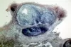 | BO_myco_skulpin_x20_01.jpg | All Pathogens | sculpin (unspecified) |
 | BO_Ich_x63_11.jpg | All Pathogens | |
 | BO_Ich_x63_10.jpg | All Pathogens, Fact Sheet Images | |
 | MXX29_024a_x100sybr_1h.jpg | All Pathogens, Fact Sheet Images | |
 | BO_myco_skulpin_x20_02.jpg | All Pathogens | sculpin (unspecified) |
 | BO_Ich_x63_09.jpg | All Pathogens, Fact Sheet Images | |
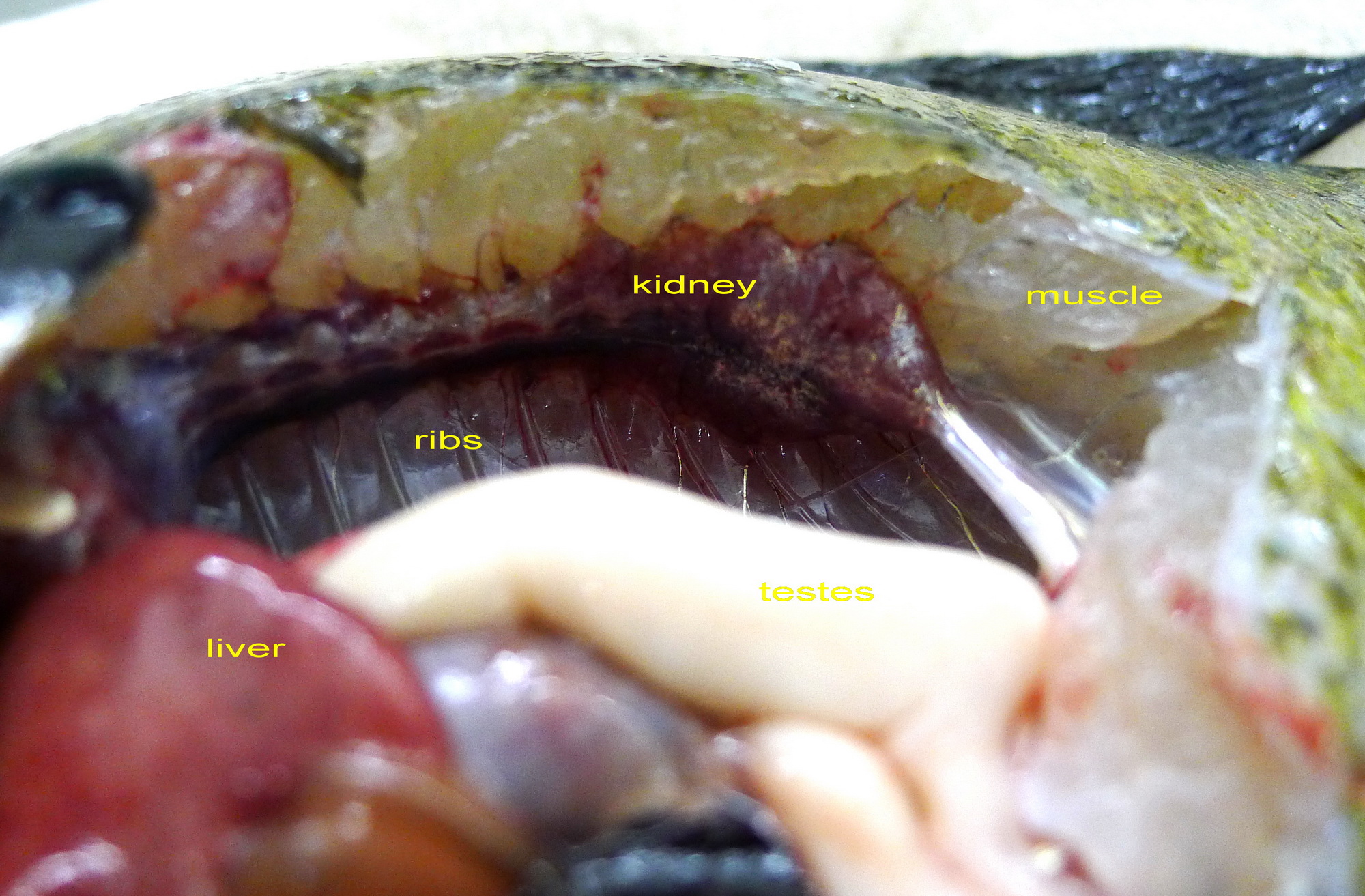
 | P1020305_annotated.jpg | All Pathogens, Fact Sheet Images | bluegill |