Skip to main content
Fish Pathogens
Login
Disclaimer
Search form
Search this site
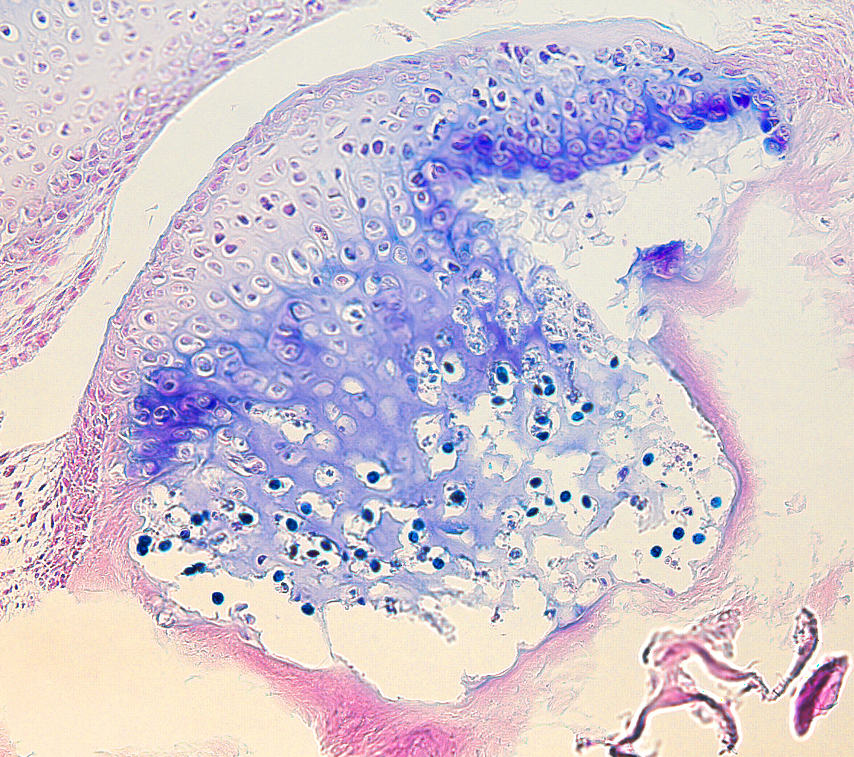
myxospore
Host:
rainbow trout
Host tissue:
head cartilage
Photographer / Illustrator:
Stephen Atkinson
Pathogen life stage:
myxospore
Image type:
Light Micrograph
Bright Field
Giemsa
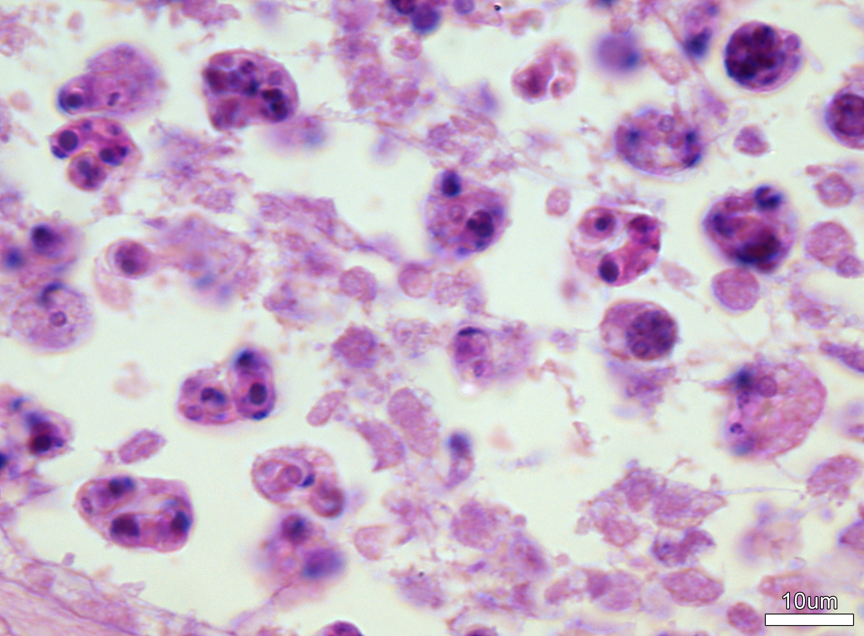
Click image to view at maximum resolution
Host tissue:
gut / intestine
Photographer / Illustrator:
Jerri Bartholomew
Pathogen life stage:
myxospore
sporoblast
Image type:
Light Micrograph
Bright Field
Giemsa
Click image to view at maximum resolution
Host tissue:
scales
Photographer / Illustrator:
Craig Banner
Pathogen life stage:
myxospore
Image type:
Light Micrograph
Bright Field
unstained
Click image to view at maximum resolution
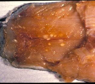
Fish muscle showing numerous white cysts of Henneguya salminicola.
Host tissue:
muscle
Photographer / Illustrator:
Craig Banner
Pathogen life stage:
cyst
myxospore
Image type:
Regular photograph
Click image to view at maximum resolution
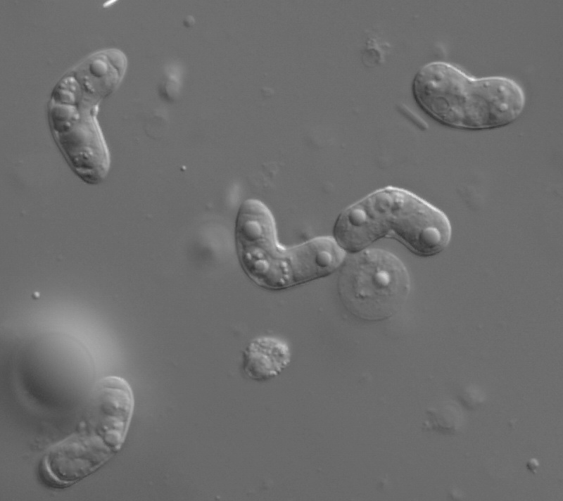
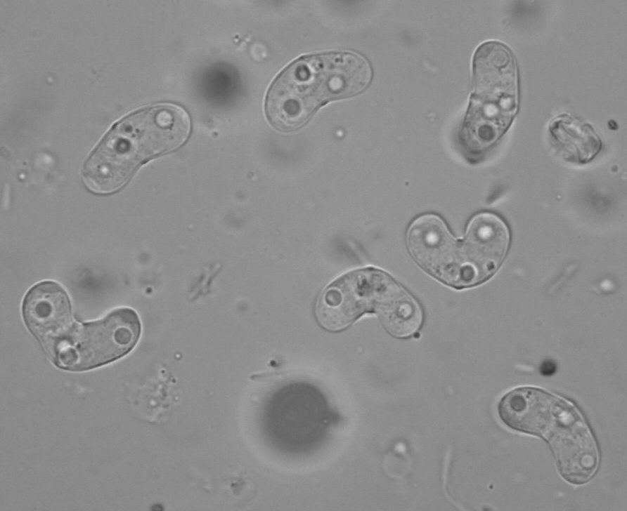
Host tissue:
n/a
Photographer / Illustrator:
Stephen Atkinson
Pathogen life stage:
myxospore
Image type:
Light Micrograph
Nomarski
Click image to view at maximum resolution
Ceratomyxa shasta myxospores from sample L14m7709_1800a.
Host tissue:
gut / intestine
Photographer / Illustrator:
Stephen Atkinson
Pathogen life stage:
myxospore
Image type:
Light Micrograph
Bright Field
Other stain
Click image to view at maximum resolution
Henneguya myxospores imaged using fluorescent microscopy and DAPI DNA stain.
Host tissue:
muscle
Photographer / Illustrator:
Stephen Atkinson
Pathogen life stage:
spore
myxospore
Image type:
Light Micrograph
Fluorescence
DAPI stain
Click image to view at maximum resolution
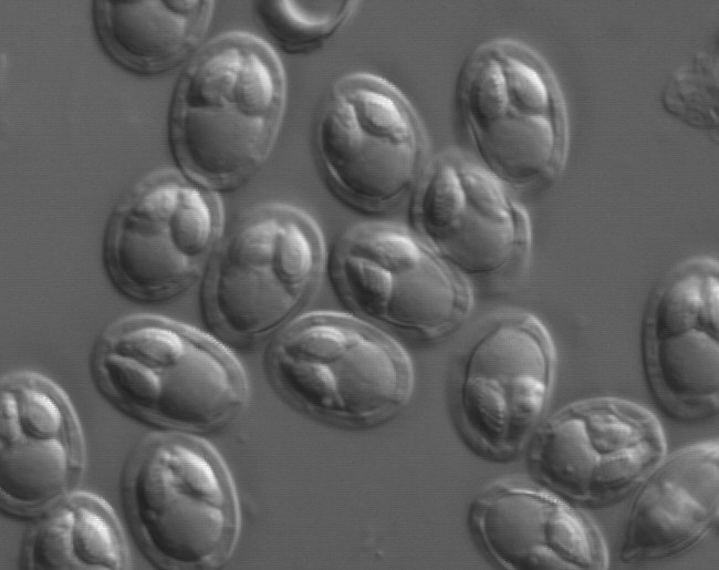
Henneguya myxospores.
Host tissue:
muscle
Photographer / Illustrator:
Stephen Atkinson
Pathogen life stage:
spore
myxospore
Image type:
Light Micrograph
Nomarski
Click image to view at maximum resolution
Myxobolus spores from the gallbladder of redside shiner.
Host:
redside shiner
Host tissue:
gall bladder
Photographer / Illustrator:
Stephen Atkinson
Pathogen life stage:
myxospore
Image type:
Light Micrograph
Nomarski
Click image to view at maximum resolution
Pages
« first
‹ previous
1
2
3