Skip to main content
Fish Pathogens
Login
Disclaimer
Search form
Search this site
myxospore
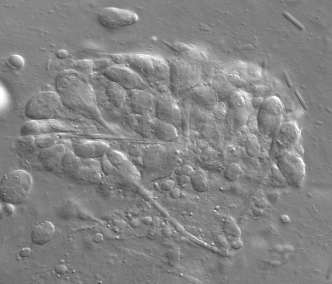
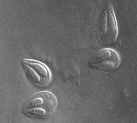
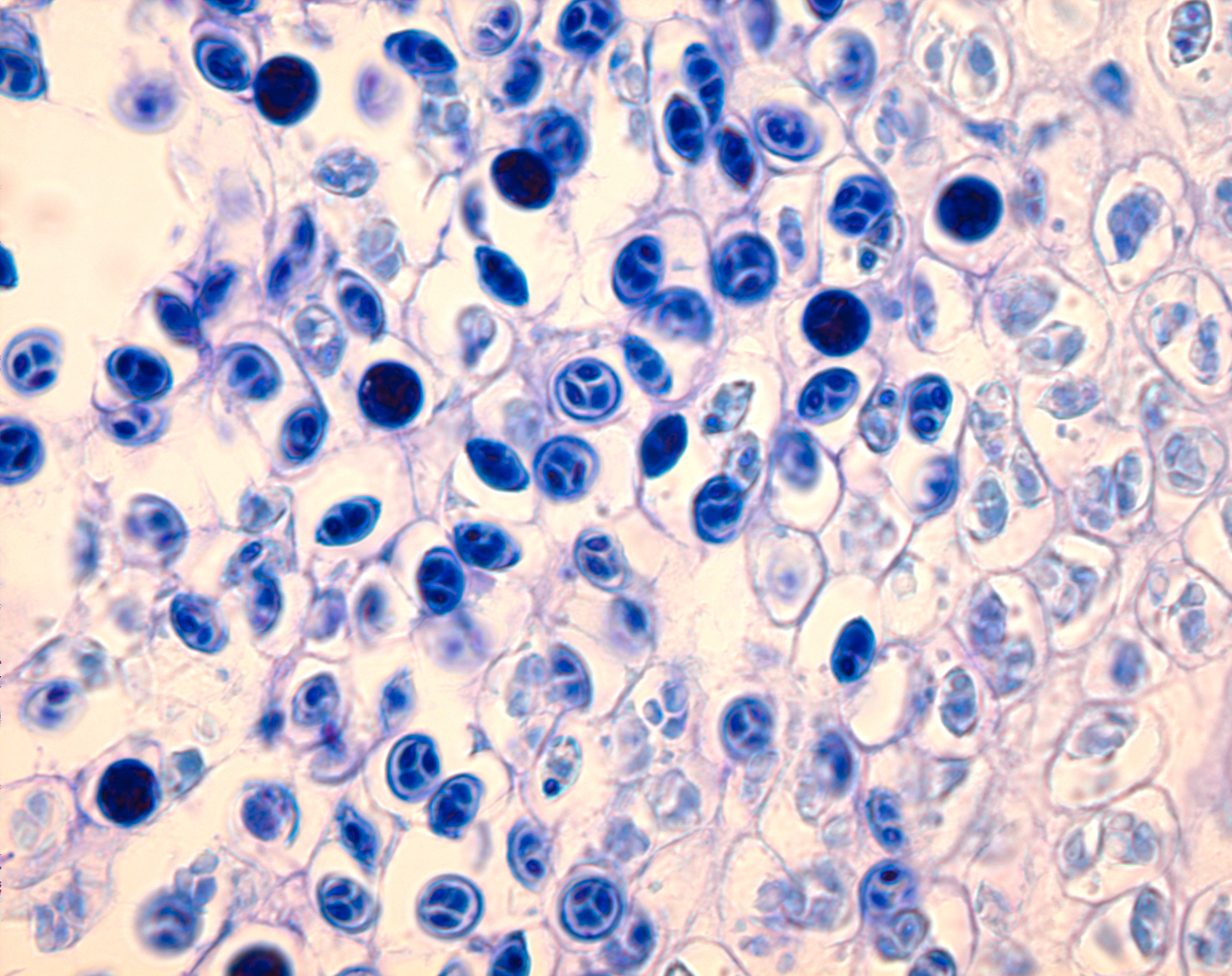
Plasmodium of developing Myxobilatus 11M21 myxospores.
Host:
largemouth bass
Host tissue:
urinary bladder
Photographer / Illustrator:
Stephen Atkinson
Pathogen life stage:
myxospore
sporoblast
Image type:
Light Micrograph
Nomarski
Click image to view at maximum resolution
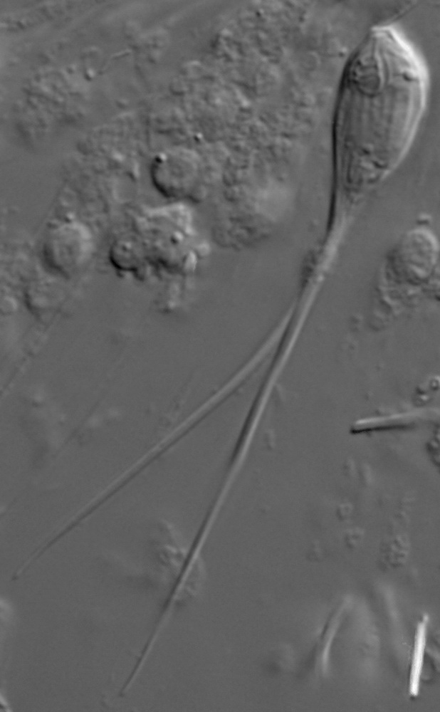
Myxospore of Myxobilatus 11M21
Host:
largemouth bass
Host tissue:
urinary bladder
Photographer / Illustrator:
Stephen Atkinson
Pathogen life stage:
myxospore
Image type:
Light Micrograph
Nomarski
Click image to view at maximum resolution
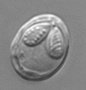
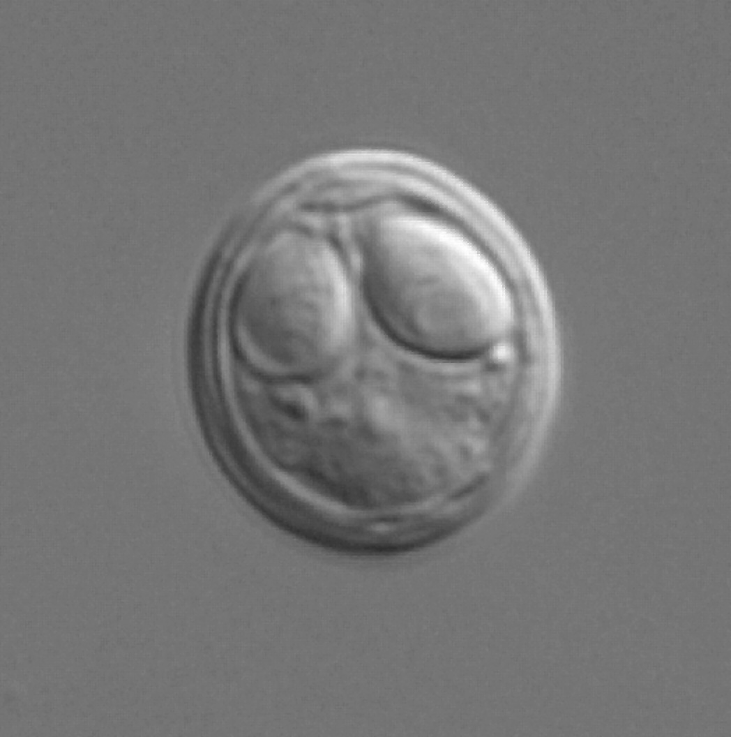
A typical Myxobolus-type myxospore.
Photographer / Illustrator:
Stephen Atkinson
Pathogen life stage:
myxospore
Image type:
Light Micrograph
Nomarski
Click image to view at maximum resolution
Life cycle of a typical myxosporean parasite.
Photographer / Illustrator:
Stephen Atkinson
Pathogen life stage:
actinospore
myxospore
Image type:
Illustration
Click image to view at maximum resolution
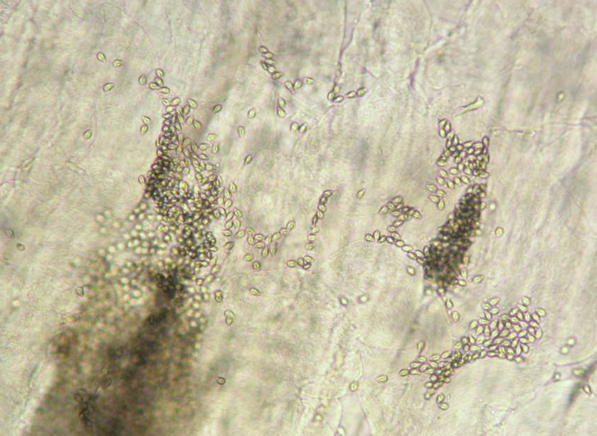
Myxobolus insidiosus myxospores.
Host tissue:
muscle
Photographer / Illustrator:
Craig Banner
Pathogen life stage:
myxospore
Image type:
Light Micrograph
Nomarski
Click image to view at maximum resolution
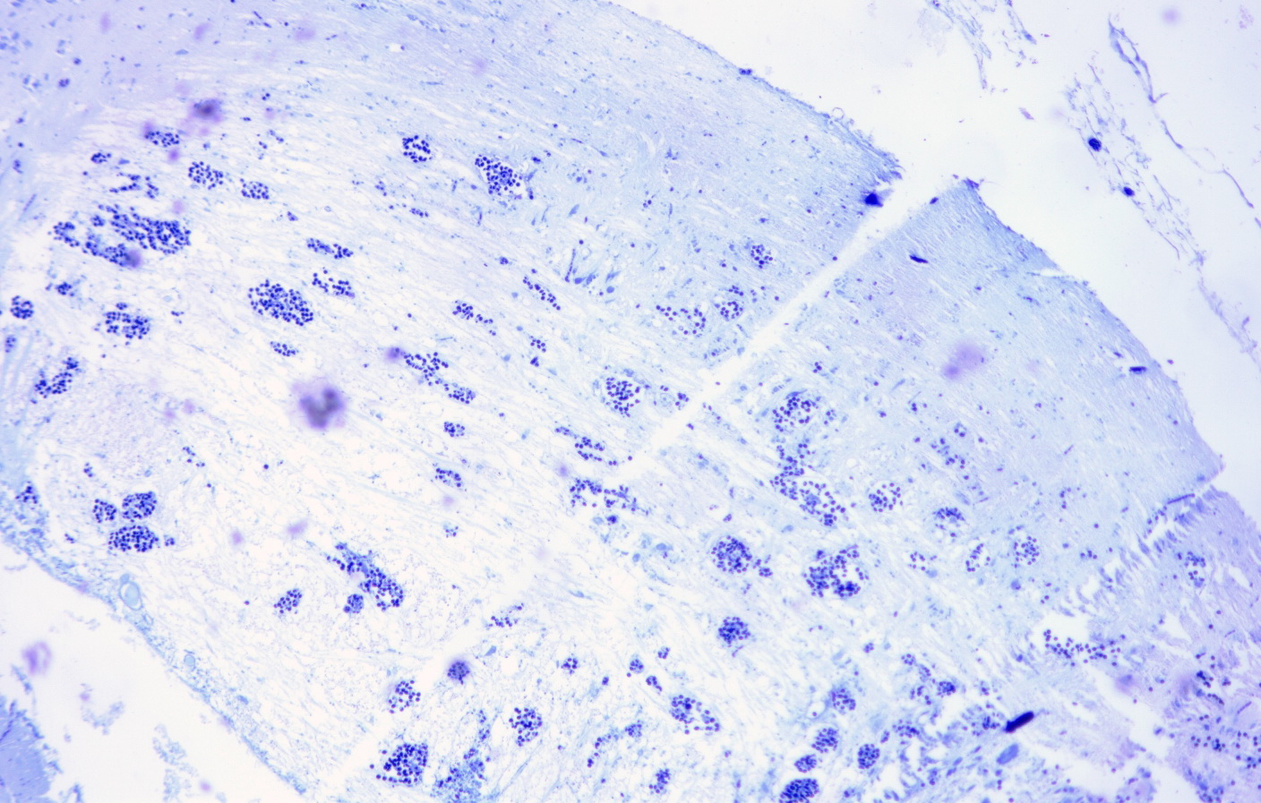
Ruptured Myxobolus insidiosus cysts in muscle.
Host tissue:
muscle
Photographer / Illustrator:
Craig Banner
Pathogen life stage:
cyst
myxospore
Image type:
Light Micrograph
Bright Field
unstained
Click image to view at maximum resolution
Host tissue:
muscle
Photographer / Illustrator:
Craig Banner
Pathogen life stage:
myxospore
Image type:
Light Micrograph
Bright Field
Giemsa
Click image to view at maximum resolution
Photographer / Illustrator:
Craig Banner
Pathogen life stage:
myxospore
Image type:
Light Micrograph
Bright Field
Giemsa
Click image to view at maximum resolution
Host:
rainbow trout
Host tissue:
n/a
Photographer / Illustrator:
Stephen Atkinson
Pathogen life stage:
myxospore
Image type:
Light Micrograph
Nomarski
Click image to view at maximum resolution
Host:
rainbow trout
Host tissue:
head cartilage
Photographer / Illustrator:
Stephen Atkinson
Pathogen life stage:
myxospore
Image type:
Light Micrograph
Bright Field
Giemsa
Click image to view at maximum resolution
Pages
« first
‹ previous
1
2
3
next ›
last »