Skip to main content
Fish Pathogens
Login
Disclaimer
Search form
Search this site
myxospore
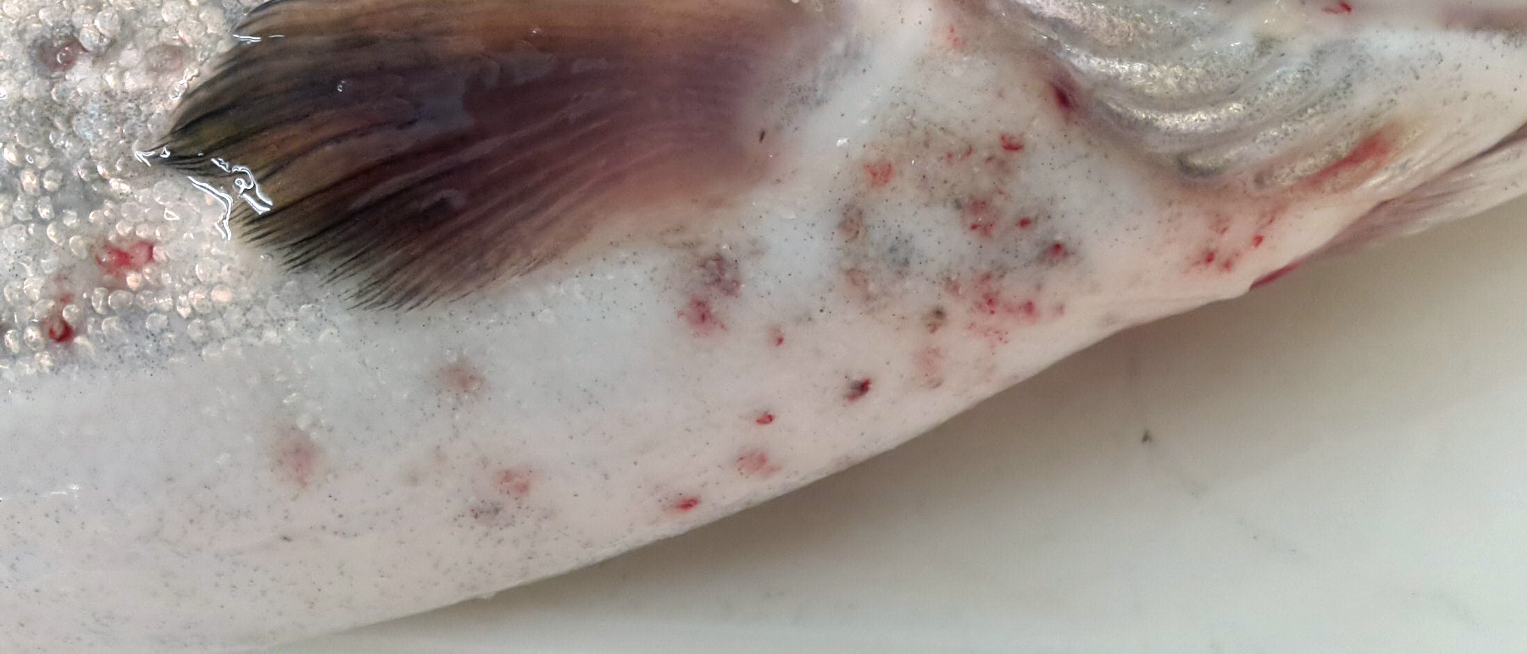
Red spots on belly of rainbow trout caused by the myxozoan parasite Myxobolus squamalis
Host:
rainbow trout
Host tissue:
scales
Photographer / Illustrator:
Stephen Atkinson
Pathogen life stage:
myxospore
Image type:
Regular photograph
Field Locality:
Leaburg Hatchery
Click image to view at maximum resolution
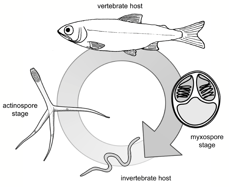
Diagram showing life cycle of Myxobolus cerebralis, with its 2 hosts and 2 spore stages.
Photographer / Illustrator:
Stephen Atkinson
Pathogen life stage:
myxospore
actinospore
Image type:
Illustration
Click image to view at maximum resolution
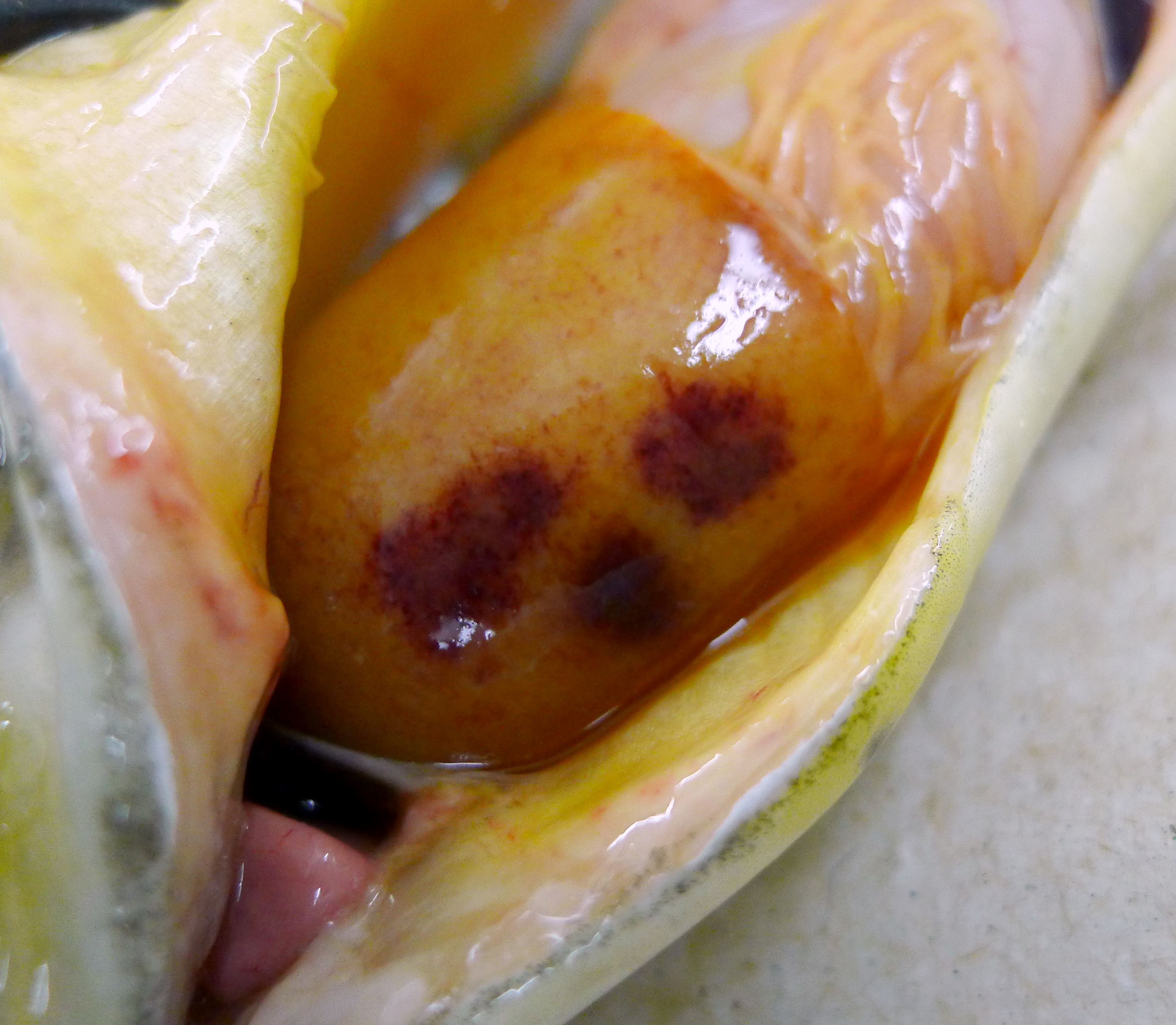
Fish liver showing dark red lesions due to C. shasta infection.
Host:
rainbow trout
Host tissue:
liver
Photographer / Illustrator:
Stephen Atkinson
Pathogen life stage:
myxospore
Image type:
Regular photograph
Click image to view at maximum resolution
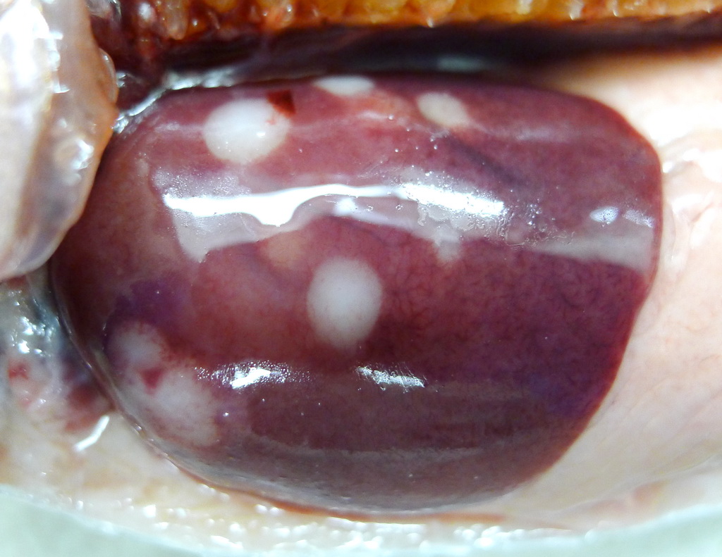
Fish liver showing pale lesions due to extensive, late-stage C. shasta infection.
Host:
rainbow trout
Host tissue:
liver
Photographer / Illustrator:
Stephen Atkinson
Pathogen life stage:
myxospore
Image type:
Regular photograph
Click image to view at maximum resolution
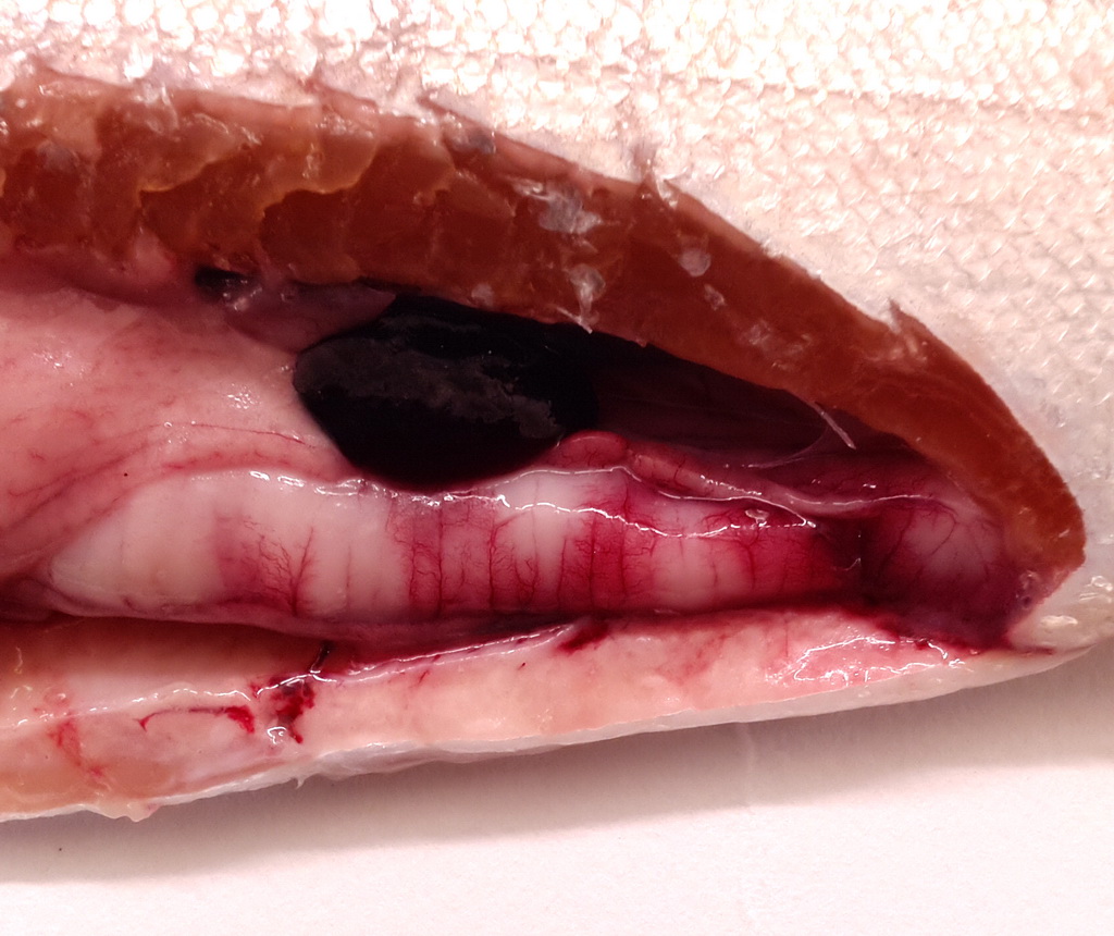
Fish intestine showing swelling and pale lesions due to extensive, late-stage C. shasta infection.
Host:
rainbow trout
Host tissue:
gut / intestine
Photographer / Illustrator:
Stephen Atkinson
Pathogen life stage:
myxospore
Image type:
Regular photograph
Click image to view at maximum resolution
Tubifex worm being placed into tube for M. cerebralis assay.
Host:
Tubifex
Host tissue:
n/a
Photographer / Illustrator:
Sascha Hallett
Pathogen life stage:
myxospore
Image type:
Regular photograph
Click image to view at maximum resolution
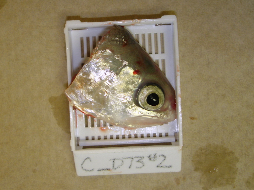
Half-head in tube prior to pepsin-trypsin digest.
Host:
rainbow trout
Host tissue:
head cartilage
Photographer / Illustrator:
Sascha Hallett
Pathogen life stage:
myxospore
Image type:
Regular photograph
Click image to view at maximum resolution
Rainbow trout half-head in histology cassette.
Host:
rainbow trout
Host tissue:
head cartilage
Photographer / Illustrator:
Sascha Hallett
Pathogen life stage:
myxospore
Image type:
Regular photograph
Click image to view at maximum resolution
Rainbow trout being sectioned for M. cerebralis detection.
Host:
rainbow trout
Host tissue:
head cartilage
Photographer / Illustrator:
Sascha Hallett
Pathogen life stage:
myxospore
Image type:
Regular photograph
Click image to view at maximum resolution
Cysts of Myxobolus spores in gill filaments.
Host tissue:
gills
Photographer / Illustrator:
Craig Banner
Pathogen life stage:
myxospore
cyst
Image type:
Light Micrograph
Bright Field
Other stain
unstained
Click image to view at maximum resolution
Pages
1
2
3
next ›
last »