 | CB_Kla_ModCWD12.jpg | All Pathogens, Fact Sheet Images | |
 | CB_CRYTO75.jpg | All Pathogens, Fact Sheet Images | |
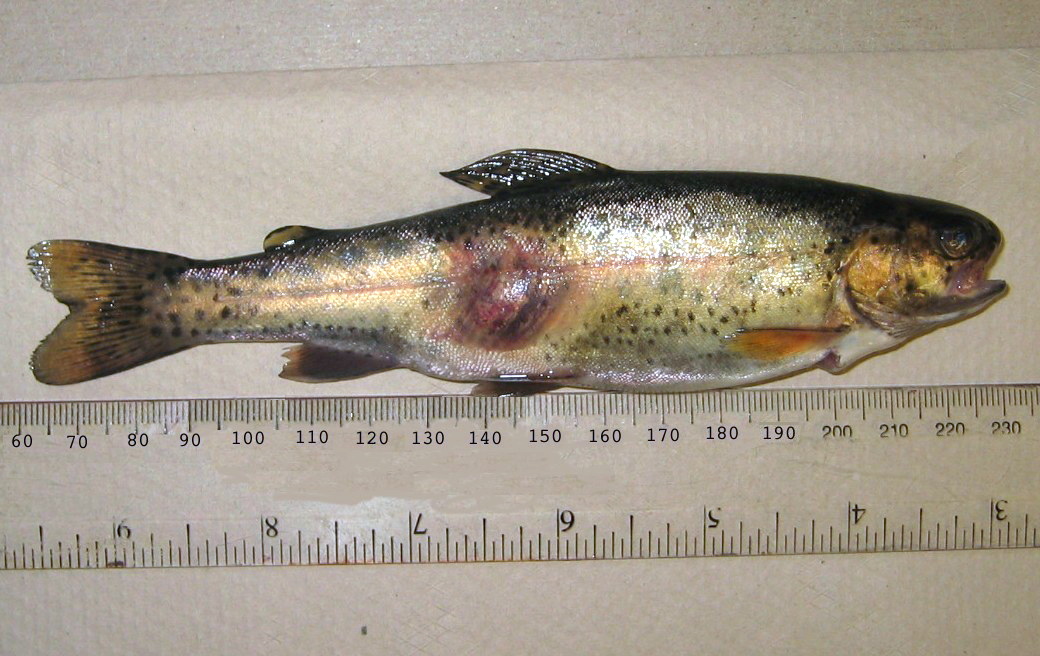
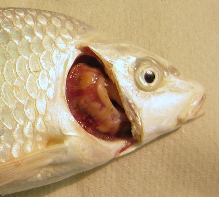
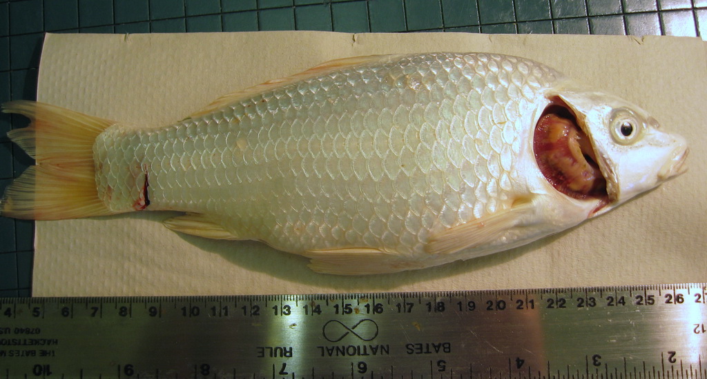
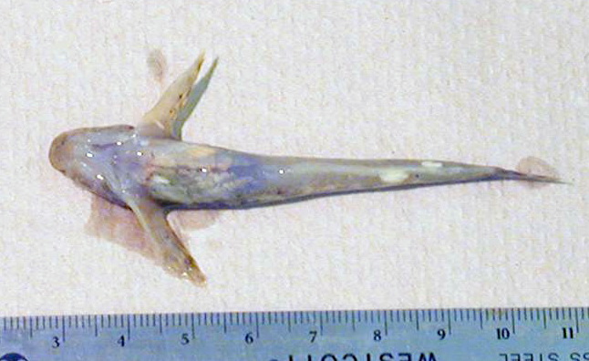
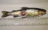
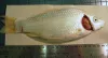
 | CB_GFFR043.jpg | All Pathogens, Fact Sheet Images | goldfish |
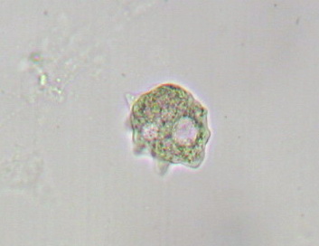
 | CB_Amoeba19.jpg | All Pathogens, Fact Sheet Images | |
 | CB_Gill_amoeb33.jpg | All Pathogens, Fact Sheet Images | |
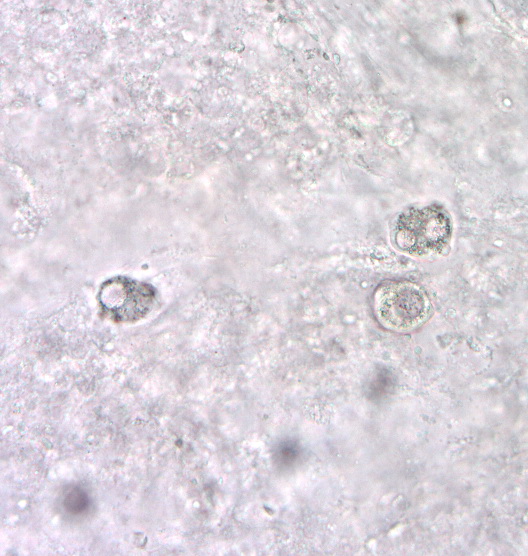
 | SA_other_para_03_x20nom_4shot_coho_kidney.jpg | All Pathogens, Fact Sheet Images | coho salmon |
 | SA_other_para_02_x20nom_4shot_coho_kidney.jpg | All Pathogens, Fact Sheet Images | coho salmon |
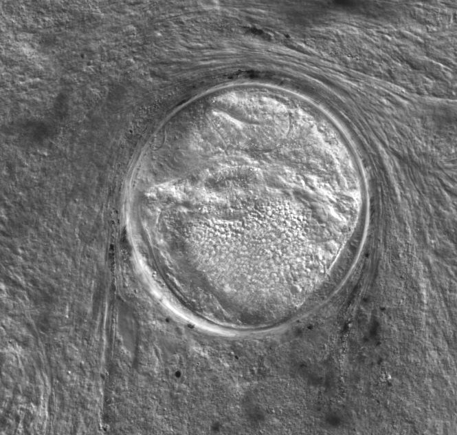
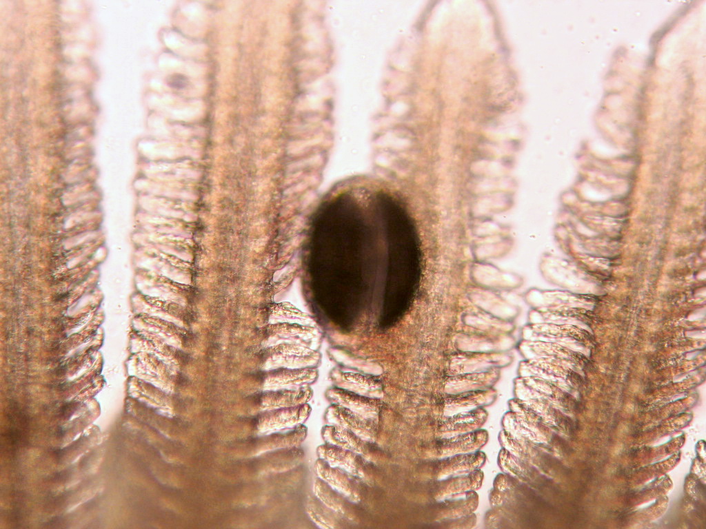
 | CB_Glochidia.jpg | All Pathogens, Fact Sheet Images | |
 | SA_other_para_01_x20nom_4shot_coho_kidney.jpg | All Pathogens, Fact Sheet Images | coho salmon |
 | SA_Loma_02b_x40bf_4shot.jpg | All Pathogens, Fact Sheet Images | |
 | CB_FCOLrc05.jpg | All Pathogens, Fact Sheet Images | |
 | SA_Loma_01a_x10bf_9shot.jpg | All Pathogens, Fact Sheet Images | |
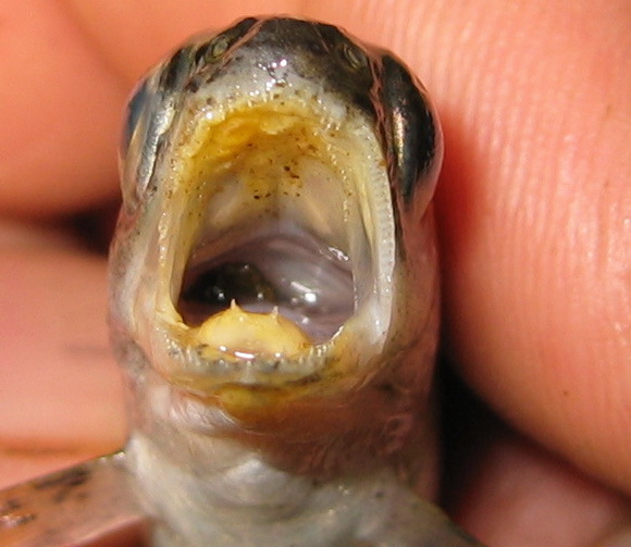
 | CB_Mouth_column4.jpg | All Pathogens, Fact Sheet Images | |
 | CB_Koi_Columnaris2b.jpg | All Pathogens, Fact Sheet Images | koi |
 | CB_Koi_Columnaris2a.jpg | All Pathogens, Hosts and other organisms, Fact Sheet Images | koi |
 | CB_MYCORASB.jpg | All Pathogens, Fact Sheet Images | |
 | CB_IMG_0448.jpg | All Pathogens, Fact Sheet Images | |
 | Minsidiosus_CB02.jpg | All Pathogens, Fact Sheet Images | |
 | Minsidiosus_CB01.jpg | All Pathogens, Fact Sheet Images | |
 | CB_IMG_0501.jpg | All Pathogens, Fact Sheet Images | |
 | Minsidiosus_CB03.jpg | All Pathogens, Fact Sheet Images | |
 | glugea_CB01.jpg | All Pathogens, Fact Sheet Images | sculpin (unspecified) |
 | CB_Myxobolus_sp5.jpg | All Pathogens, Fact Sheet Images | |
 | CB_Heterophyidae41.jpg | All Pathogens, Fact Sheet Images | |
 | CB_HeavyIch.jpg | All Pathogens, Fact Sheet Images | |
 | CB_HeavyIch2.jpg | All Pathogens, Fact Sheet Images | |
 | CB_Gtrem17.jpg | All Pathogens, Fact Sheet Images | |
 | BO_myco_skulpin_x100_04.jpg | All Pathogens | sculpin (unspecified) |
 | CB_IntTrem0.jpg | All Pathogens, Fact Sheet Images | |
 | CB_IMG_0304.jpg | All Pathogens, Fact Sheet Images | |