| P1000368ed.jpg | All Pathogens, Hosts and other organisms, Fact Sheet Images | smallmouth bass |
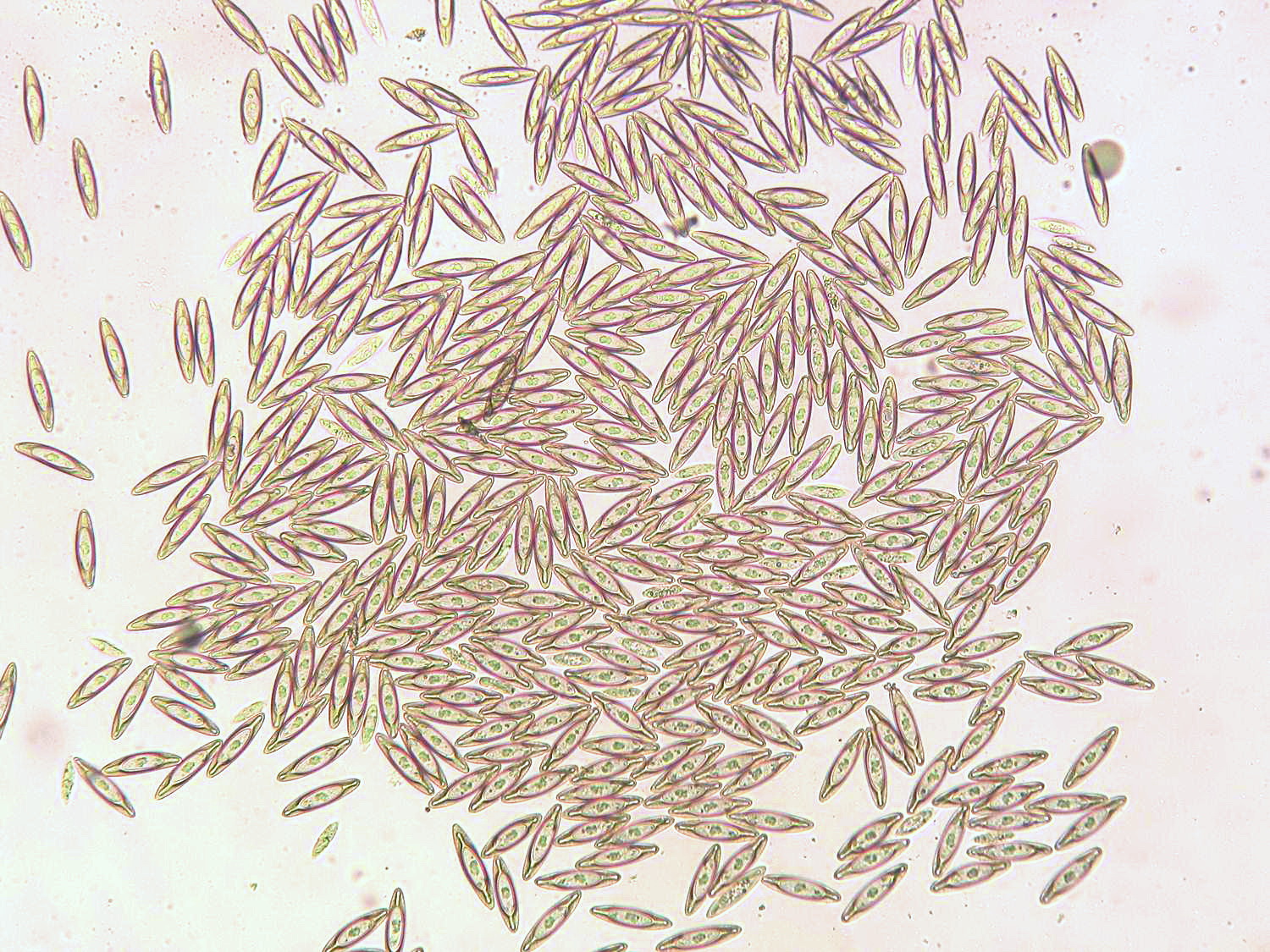
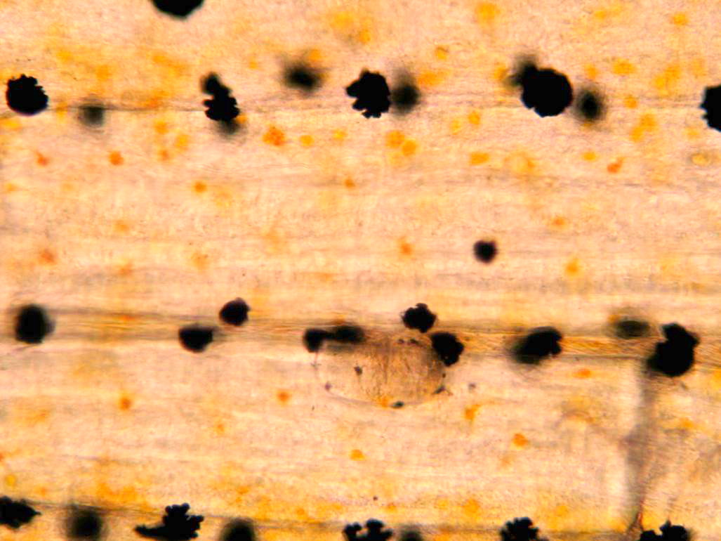
 | Acanthocephala_CB05.jpg | All Pathogens, Fact Sheet Images | |
 | Acanthocephala_CB04.jpg | All Pathogens, Fact Sheet Images | |
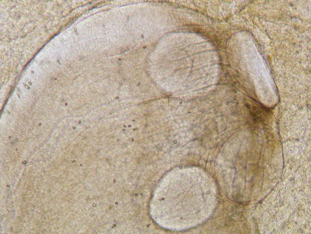
 | Acanthocephala_CB03.jpg | All Pathogens, Fact Sheet Images | |
 | P1000382ed.jpg | All Pathogens, Fact Sheet Images | smallmouth bass |
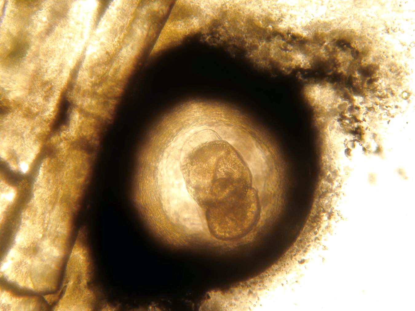
 | Acanthocephala_CB02.jpg | All Pathogens, Fact Sheet Images | |
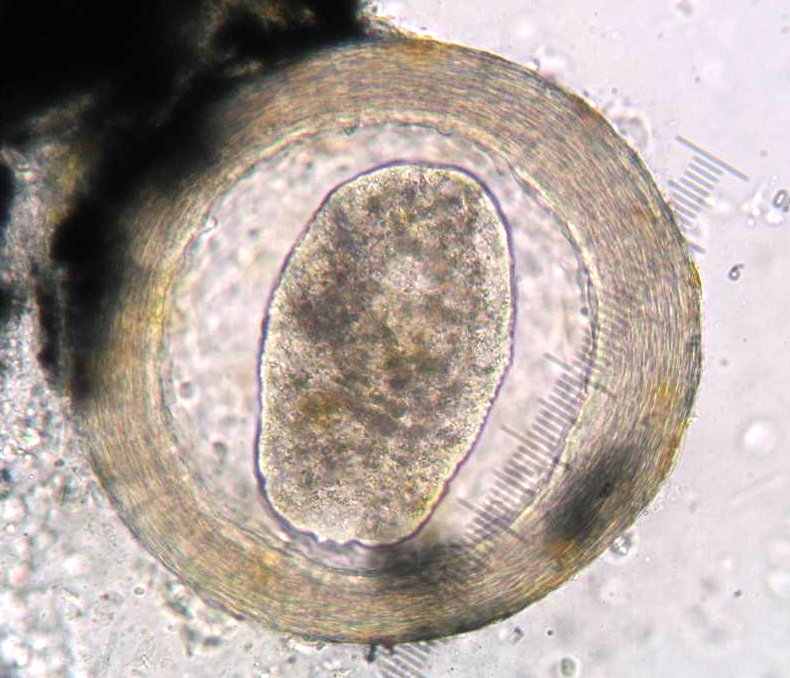
 | Acanthocephala_CB01.jpg | All Pathogens, Fact Sheet Images | |
 | P1000380ed.jpg | All Pathogens, Fact Sheet Images | smallmouth bass |
 | CB_Schistocephalus_solidus3spine.jpg | All Pathogens, Fact Sheet Images | threespine stickleback |
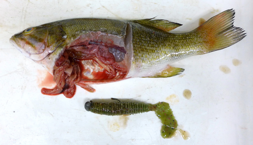
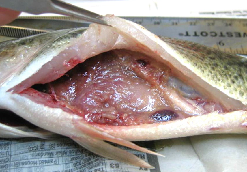
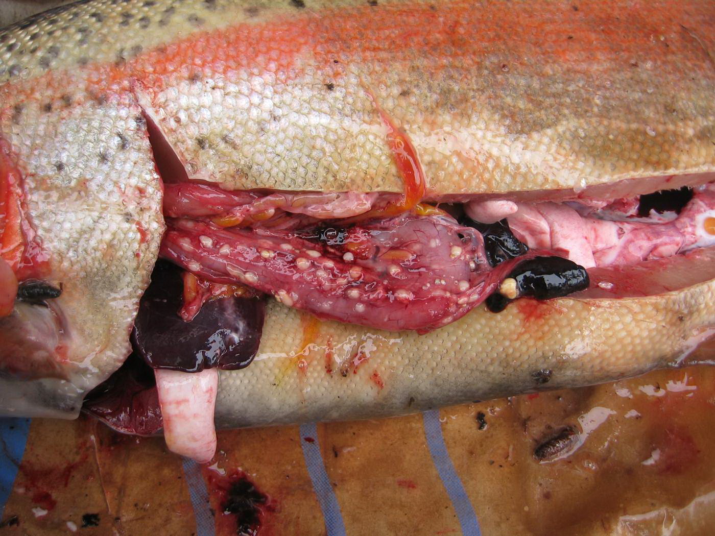
 | P1000377ed.jpg | All Pathogens, Fact Sheet Images | smallmouth bass |
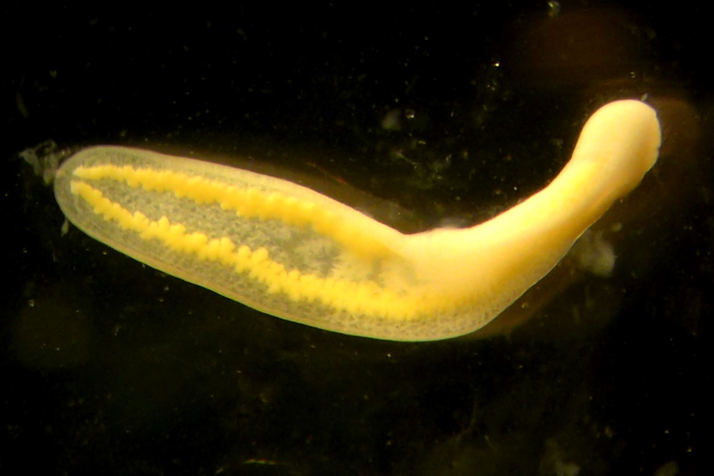
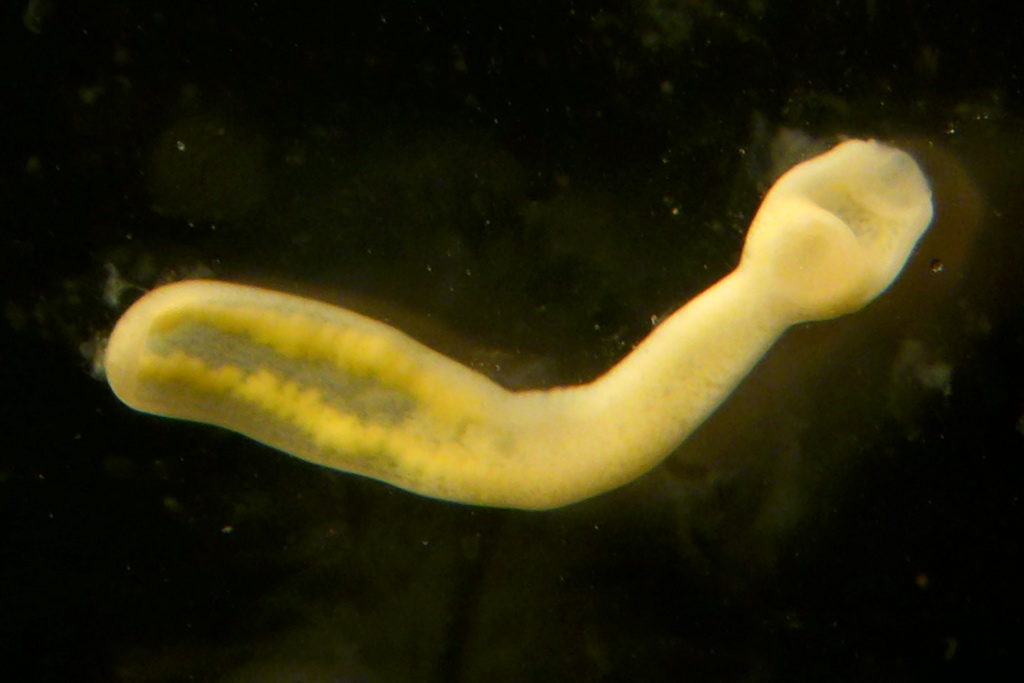
 | cestode_Lingula_intestinalis_CB01.jpg | All Pathogens, Fact Sheet Images, Top 10 | |
 | cestode_Phyllobothrium_sp_CB01.jpg | All Pathogens, Fact Sheet Images | |
 | P1000375ed.jpg | All Pathogens, Fact Sheet Images | smallmouth bass |
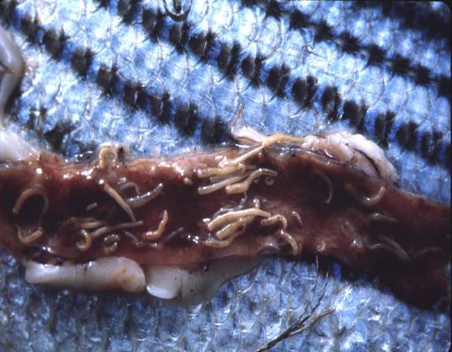
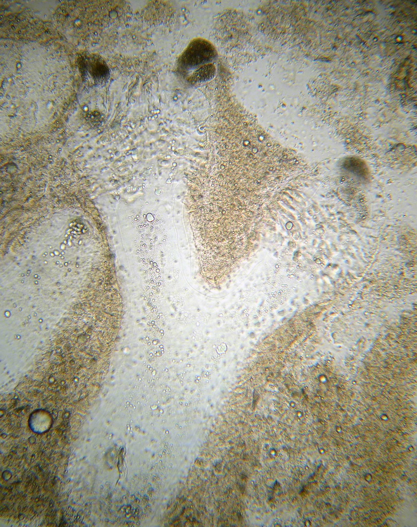
 | cesode_Proteocephalus_ambloplitis_CB01.jpg | All Pathogens, Fact Sheet Images | |
 | cesode_Proteocephalus_sp_CB02.jpg | All Pathogens, Fact Sheet Images | |
 | cesode_Bothriocephalus_sp_CB03.jpg | All Pathogens, Fact Sheet Images | |
 | Neascus_06.jpg | All Pathogens, Fact Sheet Images | |
 | Neascus_05.jpg | All Pathogens, Fact Sheet Images | |
 | Neascus_02.jpg | All Pathogens, Fact Sheet Images | |
 | nematode_Anasakis_sp_CB01.jpg | All Pathogens, Fact Sheet Images | |
 | DSC03055_deformity.jpg | All Pathogens | rainbow trout |
 | cesode_Bothriocephalus_sp_CB02.jpg | All Pathogens, Fact Sheet Images | |
 | P1000367_db.jpg | All Pathogens, Fact Sheet Images | |
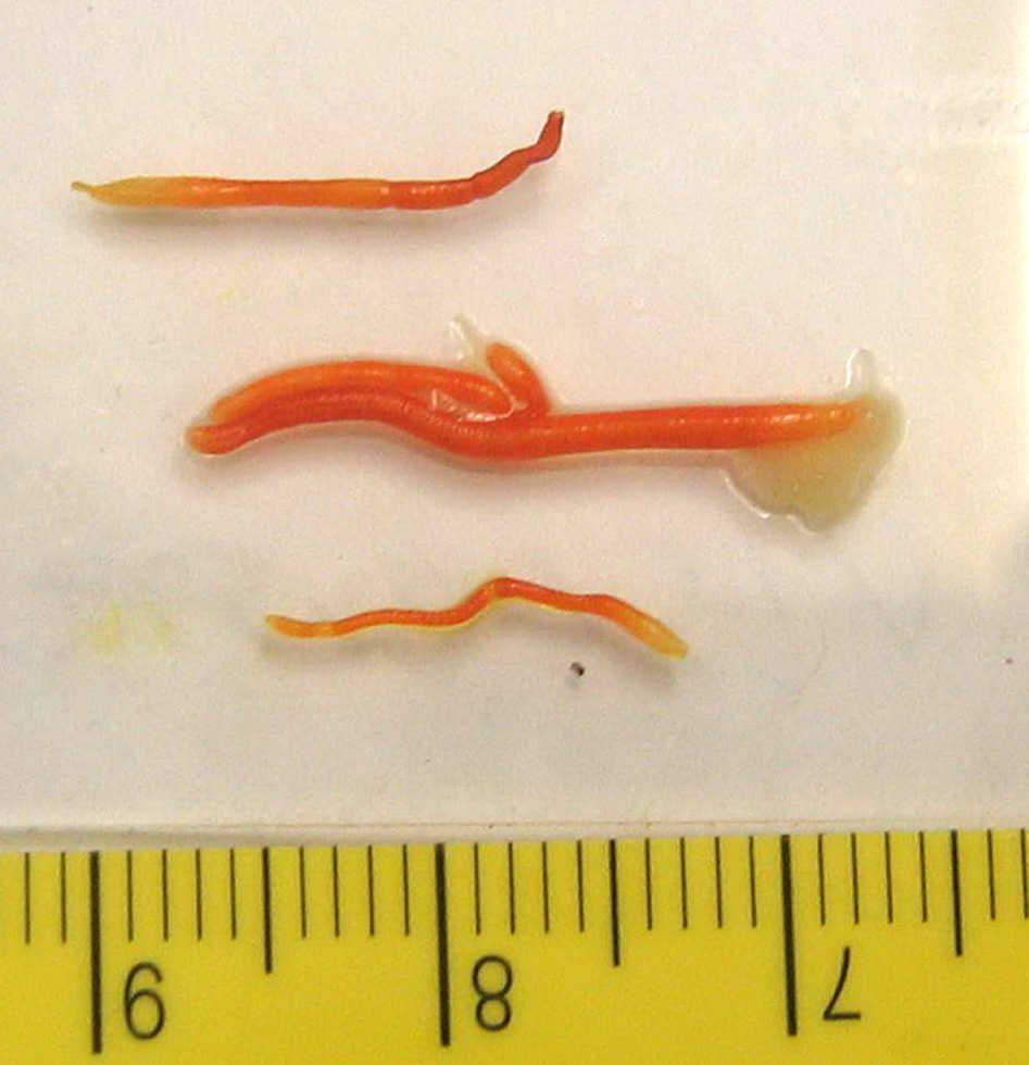
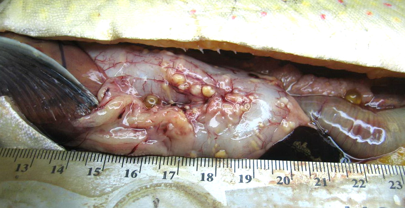
 | cesode_Diphyllobothrium_sp_CB02.jpg | All Pathogens, Fact Sheet Images | |
 | cesode_Proteocephalus_sp_CB01.jpg | All Pathogens, Fact Sheet Images | |
 | P1000365_db.jpg | All Pathogens, Fact Sheet Images | |
 | cesode_Diphyllobothrium_sp_CB03.jpg | All Pathogens, Fact Sheet Images, Top 10 | |
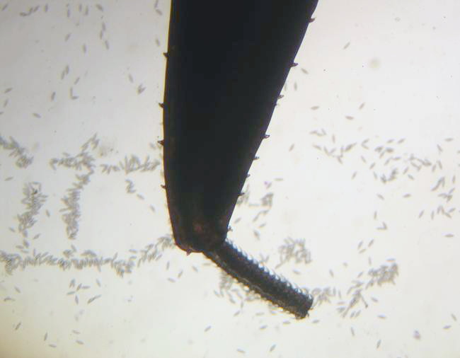
 | Acanthocephala_Echinorhynchus_gadi_CB01.jpg | All Pathogens, Fact Sheet Images | |
 | P1000366_db.jpg | All Pathogens, Fact Sheet Images | |
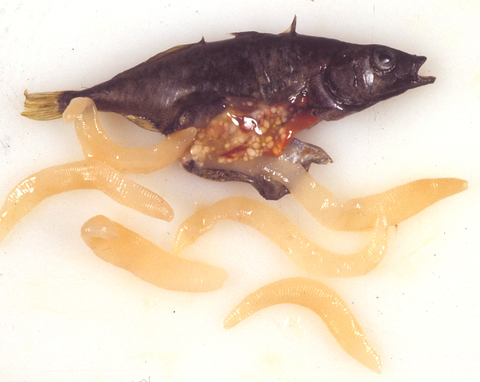
 | cesode_Diphyllobothrium_sp_CB01.jpg | All Pathogens, Fact Sheet Images | rainbow trout |